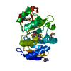
 Movie
Movie Controller


+ Open data

- Basic information
| Entry | Database: PDB / ID: 1kk9 | ||||||
|---|---|---|---|---|---|---|---|
| Title | CRYSTAL STRUCTURE OF E. COLI YCIO | ||||||
 Components Components | probable translation factor yciO | ||||||
 Keywords Keywords | UNKNOWN FUNCTION / alpha/beta open twisted sheet / Montreal-Kingston Bacterial Structural Genomics Initiative / BSGI / Structural Genomics | ||||||
| Function / homology |  Function and homology information Function and homology information | ||||||
| Biological species |  | ||||||
| Method |  X-RAY DIFFRACTION / SYNCHROTRON / MAD / Resolution: 2.1 Å X-RAY DIFFRACTION / SYNCHROTRON / MAD / Resolution: 2.1 Å | ||||||
 Authors Authors | Jia, J. / Lunin, V.V. / Sauve, V. / Huang, L.-W. / Matte, A. / Cygler, M. / Montreal-Kingston Bacterial Structural Genomics Initiative (BSGI) | ||||||
 Citation Citation | Journal: PROTEINS: STRUCT.,FUNCT.,GENET. / Year: 2002 Title: Crystal structure of the YciO protein from Escherichia coli Authors: Jia, J. / Lunin, V.V. / Sauve, V. / Huang, L.-W. / Matte, A. / Cygler, M. #1: Journal: Protein Sci. / Year: 2000Title: The structure of the Yrdc gene product from E.coli Authors: Teplova, M. / Tereshko, R. / Sanishvili, A. / Joachimiak, A. / Bushueva, T. / Anderson, W.F. / Egli, M. | ||||||
| History |
|
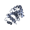
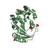
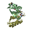
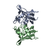
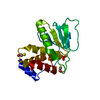
- Structure visualization
| Structure viewer | Molecule: MolmilJmol/JSmol |
|---|
- Downloads & links
-Download
| PDBx/mmCIF format | 1kk9.cif.gz | 56.7 KB | Display | PDBx/mmCIF format |
|---|---|---|---|---|
| PDB format | pdb1kk9.ent.gz | 40.4 KB | Display | PDB format |
| PDBx/mmJSON format | 1kk9.json.gz | Tree view | PDBx/mmJSON format | |
| Others |  Other downloads Other downloads |
-Validation report
| Summary document | 1kk9_validation.pdf.gz | 438 KB | Display | wwPDB validaton report |
|---|---|---|---|---|
| Full document | 1kk9_full_validation.pdf.gz | 440.7 KB | Display | |
| Data in XML | 1kk9_validation.xml.gz | 12.9 KB | Display | |
| Data in CIF | 1kk9_validation.cif.gz | 16.8 KB | Display | |
| Arichive directory | https://data.pdbj.org/pub/pdb/validation_reports/kk/1kk9ftp://data.pdbj.org/pub/pdb/validation_reports/kk/1kk9 | HTTPS FTP |
-Related structure data
| Similar structure data | |
|---|---|
| Other databases |


-Links
 PDBj
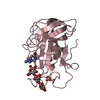
PDBj- Assembly
| Deposited unit | 
| ||||||||
|---|---|---|---|---|---|---|---|---|---|
| 1 |
| ||||||||
| Unit cell |
|
-Components
| #1: Protein | Mass: 25190.631 Da / Num. of mol.: 1 Source method: isolated from a genetically manipulated source Source: (gene. exp.) |
|---|---|
| #2: Chemical | ChemComp-SO4 /   Mass: 96.063 Da / Num. of mol.: 1 / Source method: obtained synthetically / Formula: SO4 Mass: 96.063 Da / Num. of mol.: 1 / Source method: obtained synthetically / Formula: SO4 |
| #3: Water | ChemComp-HOH /  Mass: 18.015 Da / Num. of mol.: 126 / Source method: isolated from a natural source / Formula: H2O Mass: 18.015 Da / Num. of mol.: 126 / Source method: isolated from a natural source / Formula: H2O |
| Has protein modification | Y |
-Experimental details
-Experiment
| Experiment | Method: X-RAY DIFFRACTION / Number of used crystals: 1 |
|---|
- Sample preparation
| Crystal | Density Matthews: 3.47 Å3/Da / Density % sol: 64.6 % | ||||||||||||||||||||||||||||||||||||||||||||||||||||||||||||||||||||||
|---|---|---|---|---|---|---|---|---|---|---|---|---|---|---|---|---|---|---|---|---|---|---|---|---|---|---|---|---|---|---|---|---|---|---|---|---|---|---|---|---|---|---|---|---|---|---|---|---|---|---|---|---|---|---|---|---|---|---|---|---|---|---|---|---|---|---|---|---|---|---|---|
| Crystal grow | Temperature: 298 K / Method: vapor diffusion, hanging drop / pH: 6.5 Details: PEG 3350, MES, magnesium acetate, ethylene glycol, pH 6.5, VAPOR DIFFUSION, HANGING DROP, temperature 298K | ||||||||||||||||||||||||||||||||||||||||||||||||||||||||||||||||||||||
| Crystal grow | *PLUS pH: 7.5 | ||||||||||||||||||||||||||||||||||||||||||||||||||||||||||||||||||||||
| Components of the solutions | *PLUS
|
-Data collection
| Diffraction | Mean temperature: 100 K | ||||||||||||
|---|---|---|---|---|---|---|---|---|---|---|---|---|---|
| Diffraction source | Source: SYNCHROTRON / Site: NSLS  / Beamline: X8C / Wavelength: 0.97921,0.97942,0.95369 / Beamline: X8C / Wavelength: 0.97921,0.97942,0.95369 | ||||||||||||
| Detector | Type: ADSC QUANTUM 4 / Detector: CCD / Date: Jul 2, 2001 | ||||||||||||
| Radiation | Protocol: MAD / Monochromatic (M) / Laue (L): M / Scattering type: x-ray | ||||||||||||
| Radiation wavelength |
| ||||||||||||
| Reflection | Resolution: 2.1→50 Å / Num. all: 19133 / Num. obs: 18736 / % possible obs: 99.9 % / Observed criterion σ(F): 0 / Redundancy: 10.7 % / Rsym value: 0.068 / Net I/σ(I): 10.4 | ||||||||||||
| Reflection shell | Resolution: 2.1→2.18 Å / Num. unique all: 1869 / Rsym value: 0.422 / % possible all: 100 | ||||||||||||
| Reflection | *PLUS Lowest resolution: 50 Å / Num. obs: 19133 / Num. measured all: 205748 / Rmerge(I) obs: 0.068 | ||||||||||||
| Reflection shell | *PLUS Highest resolution: 2.1 Å / % possible obs: 99.9 % / Rmerge(I) obs: 0.422 |
- Processing
| Software |
| ||||||||||||||||||||||||||||||||||||||||||||||||||||||||||||
|---|---|---|---|---|---|---|---|---|---|---|---|---|---|---|---|---|---|---|---|---|---|---|---|---|---|---|---|---|---|---|---|---|---|---|---|---|---|---|---|---|---|---|---|---|---|---|---|---|---|---|---|---|---|---|---|---|---|---|---|---|---|
| Refinement | Method to determine structure: MAD / Resolution: 2.1→50 Å / Cross valid method: THROUGHOUT / σ(F): 0 / Stereochemistry target values: Engh and Huber
| ||||||||||||||||||||||||||||||||||||||||||||||||||||||||||||
| Refinement step | Cycle: LAST / Resolution: 2.1→50 Å
| ||||||||||||||||||||||||||||||||||||||||||||||||||||||||||||
| Refine LS restraints |
| ||||||||||||||||||||||||||||||||||||||||||||||||||||||||||||
| Refinement | *PLUS Lowest resolution: 50 Å / Rfactor Rfree: 0.229 / Rfactor Rwork: 0.211 | ||||||||||||||||||||||||||||||||||||||||||||||||||||||||||||
| Solvent computation | *PLUS | ||||||||||||||||||||||||||||||||||||||||||||||||||||||||||||
| Displacement parameters | *PLUS | ||||||||||||||||||||||||||||||||||||||||||||||||||||||||||||
| Refine LS restraints | *PLUS
|
