 Movie
Movie Controller

[English] 日本語
 Yorodumi
- PDB-6t84: crystal structure of the mycobacterial trehalose monomycolate tra... -
+ Open data

- Basic information
| Entry | Database: PDB / ID: 6t84 | ||||||
|---|---|---|---|---|---|---|---|
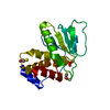
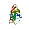
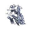
| Title | crystal structure of the mycobacterial trehalose monomycolate transport factor A, TtfA | ||||||
 Components Components | Uncharacterized protein | ||||||
 Keywords Keywords | LIPID TRANSPORT / trehalose monomycolate / MmpL3 accessory protein | ||||||
| Function / homology | : / : / cell tip / cell septum / lipid transport / cell wall organization / plasma membrane / Trehalose monomycolate transport factor A Function and homology information Function and homology information | ||||||
| Biological species |  Mycolicibacterium smegmatis MC2 155 (bacteria) Mycolicibacterium smegmatis MC2 155 (bacteria) | ||||||
| Method |  X-RAY DIFFRACTION / SYNCHROTRON / SAD / Resolution: 1.4 Å X-RAY DIFFRACTION / SYNCHROTRON / SAD / Resolution: 1.4 Å | ||||||
 Authors Authors | Blaise, M. | ||||||
| Funding support |  France, 1items France, 1items
| ||||||
 Citation Citation | Journal: Proteins / Year: 2020 Title: The crystal structure of the mycobacterial trehalose monomycolate transport factor A, TtfA, reveals an atypical fold. Authors: Ung, K.L. / Alsarraf, H.M.A.B. / Kremer, L. / Blaise, M. | ||||||
| History |
|
- Structure visualization
| Structure viewer | Molecule: MolmilJmol/JSmol |
|---|
- Downloads & links
-Download
| PDBx/mmCIF format | 6t84.cif.gz | 96.3 KB | Display | PDBx/mmCIF format |
|---|---|---|---|---|
| PDB format | pdb6t84.ent.gz | 65 KB | Display | PDB format |
| PDBx/mmJSON format | 6t84.json.gz | Tree view | PDBx/mmJSON format | |
| Others |  Other downloads Other downloads |
-Validation report
| Arichive directory | https://data.pdbj.org/pub/pdb/validation_reports/t8/6t84ftp://data.pdbj.org/pub/pdb/validation_reports/t8/6t84 | HTTPS FTP |
|---|
-Related structure data
| Similar structure data |
|---|


-Links
 PDBj
PDBj- Assembly
| Deposited unit | 
| ||||||||||||
|---|---|---|---|---|---|---|---|---|---|---|---|---|---|
| 1 |
| ||||||||||||
| Unit cell |
| ||||||||||||
| Components on special symmetry positions |
|
-Components
| #1: Protein | Mass: 21417.613 Da / Num. of mol.: 1 Source method: isolated from a genetically manipulated source Details: GHM in N-terminus is a tag as well as AS in C-terminus Source: (gene. exp.) Mycolicibacterium smegmatis MC2 155 (bacteria)Gene: MSMEG_0736 / Production host: | ||||
|---|---|---|---|---|---|
| #2: Chemical |   Mass: 96.063 Da / Num. of mol.: 2 / Source method: obtained synthetically / Formula: SO4 Mass: 96.063 Da / Num. of mol.: 2 / Source method: obtained synthetically / Formula: SO4#3: Water | ChemComp-HOH / |  Mass: 18.015 Da / Num. of mol.: 183 / Source method: isolated from a natural source / Formula: H2O Mass: 18.015 Da / Num. of mol.: 183 / Source method: isolated from a natural source / Formula: H2OHas ligand of interest | N | |
-Experimental details
-Experiment
| Experiment | Method: X-RAY DIFFRACTION / Number of used crystals: 1 |
|---|
- Sample preparation
| Crystal | Density Matthews: 2.19 Å3/Da / Density % sol: 43.83 % |
|---|---|
| Crystal grow | Temperature: 291 K / Method: vapor diffusion, sitting drop / pH: 8 / Details: 28% PEG 4000 and 0.2 M (NH4)2SO4 |
-Data collection
| Diffraction | Mean temperature: 100 K / Serial crystal experiment: N |
|---|---|
| Diffraction source | Source: SYNCHROTRON / Site: SLS  / Beamline: X06DA / Wavelength: 0.979 Å / Beamline: X06DA / Wavelength: 0.979 Å |
| Detector | Type: DECTRIS PILATUS 2M-F / Detector: PIXEL / Date: Sep 1, 2019 |
| Radiation | Protocol: SINGLE WAVELENGTH / Monochromatic (M) / Laue (L): M / Scattering type: x-ray |
| Radiation wavelength | Wavelength: 0.979 Å / Relative weight: 1 |
| Reflection | Resolution: 1.4→46.54 Å / Num. obs: 37320 / % possible obs: 99.8 % / Redundancy: 4.5 % / Biso Wilson estimate: 17.93 Å2 / CC1/2: 0.99 / Rmerge(I) obs: 0.05072 / Rpim(I) all: 0.02624 / Rrim(I) all: 0.05729 / Net I/σ(I): 14.79 |
| Reflection shell | Resolution: 1.4→1.45 Å / Rmerge(I) obs: 1.118 / Num. unique obs: 3670 / CC1/2: 0.589 |
- Processing
| Software |
| ||||||||||||||||||||||||||||||||||||||||||||||||||||||||||||||||||||||||||||||||||||||||||||||||||
|---|---|---|---|---|---|---|---|---|---|---|---|---|---|---|---|---|---|---|---|---|---|---|---|---|---|---|---|---|---|---|---|---|---|---|---|---|---|---|---|---|---|---|---|---|---|---|---|---|---|---|---|---|---|---|---|---|---|---|---|---|---|---|---|---|---|---|---|---|---|---|---|---|---|---|---|---|---|---|---|---|---|---|---|---|---|---|---|---|---|---|---|---|---|---|---|---|---|---|---|
| Refinement | Method to determine structure: SAD / Resolution: 1.4→46.54 Å / SU ML: 0.1644 / Cross valid method: FREE R-VALUE / σ(F): 1.35 / Phase error: 20.8485
| ||||||||||||||||||||||||||||||||||||||||||||||||||||||||||||||||||||||||||||||||||||||||||||||||||
| Solvent computation | Shrinkage radii: 0.9 Å / VDW probe radii: 1.11 Å | ||||||||||||||||||||||||||||||||||||||||||||||||||||||||||||||||||||||||||||||||||||||||||||||||||
| Displacement parameters | Biso mean: 28.14 Å2 | ||||||||||||||||||||||||||||||||||||||||||||||||||||||||||||||||||||||||||||||||||||||||||||||||||
| Refinement step | Cycle: LAST / Resolution: 1.4→46.54 Å
| ||||||||||||||||||||||||||||||||||||||||||||||||||||||||||||||||||||||||||||||||||||||||||||||||||
| Refine LS restraints |
| ||||||||||||||||||||||||||||||||||||||||||||||||||||||||||||||||||||||||||||||||||||||||||||||||||
| LS refinement shell |
|
