 Movie
Movie Controller

[English] 日本語
 Yorodumi
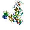
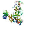
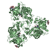
- PDB-3b18: Rv0098 of Mycobacterium tuberculosis with ordered loop between be... -
+ Open data

- Basic information
| Entry | Database: PDB / ID: 3b18 | ||||||
|---|---|---|---|---|---|---|---|
| Title | Rv0098 of Mycobacterium tuberculosis with ordered loop between beta-4 and beta-5 | ||||||
 Components Components | Uncharacterized protein Rv0098/MT0107 | ||||||
 Keywords Keywords | HYDROLASE / hot dog fold / long-chain fatty acyl-CoA thioesterase / acyl carrier protein / Mycobacterium tuberculosis | ||||||
| Function / homology |  Function and homology information Function and homology information(3R)-3-[(carboxylmethyl)amino]fatty acid synthase / palmitoyl-CoA hydrolase / fatty acyl-CoA hydrolase activity / Hydrolases; Acting on ester bonds; Thioester hydrolases / fatty acid metabolic process / fatty acid biosynthetic process / lyase activity / plasma membrane Similarity search - Function | ||||||
| Biological species |   Mycobacterium tuberculosis (bacteria) Mycobacterium tuberculosis (bacteria) | ||||||
| Method |  X-RAY DIFFRACTION / MOLECULAR REPLACEMENT / Resolution: 2.75 Å X-RAY DIFFRACTION / MOLECULAR REPLACEMENT / Resolution: 2.75 Å | ||||||
 Authors Authors | Maity, K. / Suguna, K. | ||||||
 Citation Citation | Journal: J.Biomol.Struct.Dyn. / Year: 2012 Title: Insights into the substrate specificity of a thioesterase Rv0098 of mycobacterium tuberculosis through X-ray crystallographic and molecular dynamics studies. Authors: Maity, K. / Bajaj, P. / Surolia, N. / Surolia, A. / Suguna, K. | ||||||
| History |
|
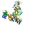
- Structure visualization
| Structure viewer | Molecule: MolmilJmol/JSmol |
|---|
- Downloads & links
-Download
| PDBx/mmCIF format | 3b18.cif.gz | 47.5 KB | Display | PDBx/mmCIF format |
|---|---|---|---|---|
| PDB format | pdb3b18.ent.gz | 33.6 KB | Display | PDB format |
| PDBx/mmJSON format | 3b18.json.gz | Tree view | PDBx/mmJSON format | |
| Others |  Other downloads Other downloads |
-Validation report
| Arichive directory | https://data.pdbj.org/pub/pdb/validation_reports/b1/3b18ftp://data.pdbj.org/pub/pdb/validation_reports/b1/3b18 | HTTPS FTP |
|---|
-Related structure data
| Related structure data | 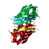 2pfcS S: Starting model for refinement |
|---|---|
| Similar structure data |
-Links
 PDBj
PDBj
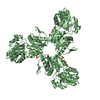
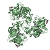
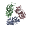
- Assembly
| Deposited unit | 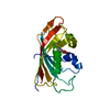
| ||||||||
|---|---|---|---|---|---|---|---|---|---|
| 1 | x 6
| ||||||||
| 2 | 
| ||||||||
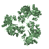
| Unit cell |
|
-Components
| #1: Protein | Mass: 20549.479 Da / Num. of mol.: 1 Source method: isolated from a genetically manipulated source Source: (gene. exp.) Mycobacterium tuberculosis (bacteria) / Gene: Rv0098 / Plasmid: PET-28a(+)(NOVAGEN) / Production host: |
|---|---|
| #2: Chemical | ChemComp-DAO /   Mass: 200.318 Da / Num. of mol.: 1 / Source method: obtained synthetically / Formula: C12H24O2 Mass: 200.318 Da / Num. of mol.: 1 / Source method: obtained synthetically / Formula: C12H24O2 |
| #3: Water | ChemComp-HOH /  Mass: 18.015 Da / Num. of mol.: 28 / Source method: isolated from a natural source / Formula: H2O Mass: 18.015 Da / Num. of mol.: 28 / Source method: isolated from a natural source / Formula: H2O |
| Has protein modification | Y |
-Experimental details
-Experiment
| Experiment | Method: X-RAY DIFFRACTION / Number of used crystals: 1 |
|---|
- Sample preparation
| Crystal | Density Matthews: 2.04 Å3/Da / Density % sol: 39.68 % |
|---|---|
| Crystal grow | Temperature: 289 K / Method: vapor diffusion, hanging drop / pH: 7.5 Details: 5%-12% PEG 6000, 0.1M HEPES buffer, 5% MPD , pH 7.5, VAPOR DIFFUSION, HANGING DROP, temperature 289K |
-Data collection
| Diffraction | Mean temperature: 100 K |
|---|---|
| Diffraction source | Source: ROTATING ANODE / Type: RIGAKU RU200 / Wavelength: 1.54 Å |
| Detector | Type: MAR scanner 345 mm plate / Detector: IMAGE PLATE / Date: Sep 15, 2006 / Details: Mirror |
| Radiation | Monochromator: Osmic Mirror / Protocol: SINGLE WAVELENGTH / Monochromatic (M) / Laue (L): M / Scattering type: x-ray |
| Radiation wavelength | Wavelength: 1.54 Å / Relative weight: 1 |
| Reflection | Resolution: 2.75→50.1 Å / Num. obs: 4502 / % possible obs: 100 % / Observed criterion σ(I): 0 / Redundancy: 16.1 % / Biso Wilson estimate: 75.7 Å2 / Rmerge(I) obs: 0.091 / Net I/σ(I): 23.3 |
| Reflection shell | Resolution: 2.75→2.9 Å / Redundancy: 16.3 % / Rmerge(I) obs: 0.76 / Mean I/σ(I) obs: 4 / Num. unique all: 648 / % possible all: 100 |
- Processing
| Software |
| |||||||||||||||||||||||||||||||||||||||||||||||||||||||||||||||||
|---|---|---|---|---|---|---|---|---|---|---|---|---|---|---|---|---|---|---|---|---|---|---|---|---|---|---|---|---|---|---|---|---|---|---|---|---|---|---|---|---|---|---|---|---|---|---|---|---|---|---|---|---|---|---|---|---|---|---|---|---|---|---|---|---|---|---|
| Refinement | Method to determine structure: MOLECULAR REPLACEMENT Starting model: 2PFC Resolution: 2.75→50.01 Å / Cor.coef. Fo:Fc: 0.93 / Cor.coef. Fo:Fc free: 0.913 / SU B: 12.006 / SU ML: 0.239 / Isotropic thermal model: RESTRAINED / Cross valid method: THROUGHOUT / σ(F): 0 / ESU R Free: 0.093 / Stereochemistry target values: MAXIMUM LIKELIHOOD / Details: BULK SOLVENT MODEL USED
| |||||||||||||||||||||||||||||||||||||||||||||||||||||||||||||||||
| Solvent computation | Ion probe radii: 0.8 Å / Shrinkage radii: 0.8 Å / VDW probe radii: 1.4 Å / Solvent model: BABINET MODEL WITH MASK | |||||||||||||||||||||||||||||||||||||||||||||||||||||||||||||||||
| Displacement parameters | Biso mean: 59.096 Å2 | |||||||||||||||||||||||||||||||||||||||||||||||||||||||||||||||||
| Refinement step | Cycle: LAST / Resolution: 2.75→50.01 Å
| |||||||||||||||||||||||||||||||||||||||||||||||||||||||||||||||||
| Refine LS restraints |
| |||||||||||||||||||||||||||||||||||||||||||||||||||||||||||||||||
| LS refinement shell | Resolution: 2.752→2.823 Å / Total num. of bins used: 20
|
