 Movie
Movie Controller

[English] 日本語
 Yorodumi
- PDB-1kgz: Crystal Structure Analysis of the Anthranilate Phosphoribosyltran... -
+ Open data

- Basic information
| Entry | Database: PDB / ID: 1kgz | ||||||
|---|---|---|---|---|---|---|---|
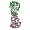
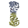
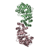
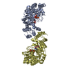
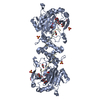
| Title | Crystal Structure Analysis of the Anthranilate Phosphoribosyltransferase from Erwinia carotovora (current name, Pectobacterium carotovorum) | ||||||
 Components Components | Anthranilate phosphoribosyltransferase | ||||||
 Keywords Keywords | TRANSFERASE / type 3 prt fold / nucleotide binding fold | ||||||
| Function / homology |  Function and homology information Function and homology informationanthranilate phosphoribosyltransferase / anthranilate phosphoribosyltransferase activity / L-tryptophan biosynthetic process / magnesium ion binding / cytosol Similarity search - Function | ||||||
| Biological species |  Pectobacterium carotovorum (bacteria) Pectobacterium carotovorum (bacteria) | ||||||
| Method |  X-RAY DIFFRACTION / MIR / Resolution: 2.4 Å X-RAY DIFFRACTION / MIR / Resolution: 2.4 Å | ||||||
 Authors Authors | Kim, C. / Xuong, N.-H. / Edwards, S. / Madhusudan / Yee, M.-C. / Spraggon, G. / Mills, S.E. | ||||||
 Citation Citation | Journal: FEBS Lett. / Year: 2002 Title: The Crystal Structure of Anthranilate Phosphoribosyltransferase from the Enterobacterium Pectobacterium carotovorum Authors: Kim, C. / Xuong, N.-H. / Edwards, S. / Madhusudan / Yee, M.-C. / Spraggon, G. / Mills, S.E. | ||||||
| History |
|
- Structure visualization
| Structure viewer | Molecule: MolmilJmol/JSmol |
|---|
- Downloads & links
-Download
| PDBx/mmCIF format | 1kgz.cif.gz | 138.2 KB | Display | PDBx/mmCIF format |
|---|---|---|---|---|
| PDB format | pdb1kgz.ent.gz | 109.5 KB | Display | PDB format |
| PDBx/mmJSON format | 1kgz.json.gz | Tree view | PDBx/mmJSON format | |
| Others |  Other downloads Other downloads |
-Validation report
| Arichive directory | https://data.pdbj.org/pub/pdb/validation_reports/kg/1kgzftp://data.pdbj.org/pub/pdb/validation_reports/kg/1kgz | HTTPS FTP |
|---|
-Related structure data


-Links
 PDBj
PDBj


- Assembly
| Deposited unit | 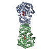
| ||||||||
|---|---|---|---|---|---|---|---|---|---|
| 1 |
| ||||||||
| Unit cell |
|
-Components
| #1: Protein | Mass: 37057.117 Da / Num. of mol.: 2 Source method: isolated from a genetically manipulated source Source: (gene. exp.) Pectobacterium carotovorum (bacteria) / Gene: trpD / Plasmid: pUC13 / Production host: References: UniProt: Q8VP84, anthranilate phosphoribosyltransferase #2: Chemical |   Mass: 200.590 Da / Num. of mol.: 2 / Source method: obtained synthetically / Formula: Hg Mass: 200.590 Da / Num. of mol.: 2 / Source method: obtained synthetically / Formula: Hg#3: Chemical | ChemComp-MN /   Mass: 54.938 Da / Num. of mol.: 4 / Source method: obtained synthetically / Formula: Mn Mass: 54.938 Da / Num. of mol.: 4 / Source method: obtained synthetically / Formula: Mn#4: Sugar |   Type: D-saccharide / Mass: 390.070 Da / Num. of mol.: 2 / Source method: obtained synthetically / Formula: C5H13O14P3 Type: D-saccharide / Mass: 390.070 Da / Num. of mol.: 2 / Source method: obtained synthetically / Formula: C5H13O14P3#5: Water | ChemComp-HOH / |  Mass: 18.015 Da / Num. of mol.: 202 / Source method: isolated from a natural source / Formula: H2O Mass: 18.015 Da / Num. of mol.: 202 / Source method: isolated from a natural source / Formula: H2O |
|---|
-Experimental details
-Experiment
| Experiment | Method: X-RAY DIFFRACTION / Number of used crystals: 1 |
|---|
- Sample preparation
| Crystal | Density Matthews: 2.17 Å3/Da / Density % sol: 43.23 % | ||||||||||||||||||||||||||||||||||||||||||||||||||||||||||||||||||||||||||||||||||||||||||||||||||
|---|---|---|---|---|---|---|---|---|---|---|---|---|---|---|---|---|---|---|---|---|---|---|---|---|---|---|---|---|---|---|---|---|---|---|---|---|---|---|---|---|---|---|---|---|---|---|---|---|---|---|---|---|---|---|---|---|---|---|---|---|---|---|---|---|---|---|---|---|---|---|---|---|---|---|---|---|---|---|---|---|---|---|---|---|---|---|---|---|---|---|---|---|---|---|---|---|---|---|---|
| Crystal grow | Temperature: 298 K / Method: vapor diffusion, hanging drop / pH: 6.5 Details: PEG 3000, MnCl2, glutamine, pH 6.5, VAPOR DIFFUSION, HANGING DROP, temperature 298K | ||||||||||||||||||||||||||||||||||||||||||||||||||||||||||||||||||||||||||||||||||||||||||||||||||
| Crystal grow | *PLUS | ||||||||||||||||||||||||||||||||||||||||||||||||||||||||||||||||||||||||||||||||||||||||||||||||||
| Components of the solutions | *PLUS
|
-Data collection
| Diffraction | Mean temperature: 200 K |
|---|---|
| Diffraction source | Source: ROTATING ANODE |
| Detector | Detector: AREA DETECTOR / Date: Aug 7, 2000 |
| Radiation | Monochromator: YALE MIRRORS / Protocol: SINGLE WAVELENGTH / Monochromatic (M) / Laue (L): M / Scattering type: x-ray |
| Radiation wavelength | Relative weight: 1 |
| Reflection | Resolution: 2.4→25 Å / Num. all: 25162 / Num. obs: 24349 / % possible obs: 98.6 % / Observed criterion σ(F): 1 / Observed criterion σ(I): 1 / Redundancy: 25 % / Rmerge(I) obs: 0.074 / Rsym value: 0.078 / Net I/σ(I): 37.6 |
| Reflection shell | Resolution: 2.4→2.49 Å / Rmerge(I) obs: 0.334 / Mean I/σ(I) obs: 7.1 / Num. unique all: 2394 / Rsym value: 0.311 / % possible all: 98.6 |
| Reflection | *PLUS Lowest resolution: 50 Å / Num. obs: 20457 / % possible obs: 98.7 % / Redundancy: 3.6 % / Rmerge(I) obs: 0.067 |
| Reflection shell | *PLUS Rmerge(I) obs: 0.398 |
- Processing
| Software |
| ||||||||||||||||||||||||
|---|---|---|---|---|---|---|---|---|---|---|---|---|---|---|---|---|---|---|---|---|---|---|---|---|---|
| Refinement | Method to determine structure: MIR / Resolution: 2.4→25 Å / Cross valid method: THROUGHOUT / σ(F): 2 / σ(I): 2 / Stereochemistry target values: Engh & Huber
| ||||||||||||||||||||||||
| Refinement step | Cycle: LAST / Resolution: 2.4→25 Å
| ||||||||||||||||||||||||
| Refinement | *PLUS Highest resolution: 2.44 Å / Lowest resolution: 50 Å / Rfactor Rfree: 0.2643 / Rfactor Rwork: 0.2125 | ||||||||||||||||||||||||
| Solvent computation | *PLUS | ||||||||||||||||||||||||
| Displacement parameters | *PLUS | ||||||||||||||||||||||||
| Refine LS restraints | *PLUS
|
