 Movie
Movie Controller

[English] 日本語
 Yorodumi
- PDB-1kbj: Crystallographic Study of the Recombinant Flavin-binding Domain o... -
+ Open data

- Basic information
| Entry | Database: PDB / ID: 1kbj | ||||||
|---|---|---|---|---|---|---|---|
| Title | Crystallographic Study of the Recombinant Flavin-binding Domain of Baker's Yeast Flavocytochrome b2: comparison with the Intact Wild-type Enzyme | ||||||
 Components Components | CYTOCHROME B2 | ||||||
 Keywords Keywords | OXIDOREDUCTASE / flavin-binding domain of flavocytochrome b2 | ||||||
| Function / homology |  Function and homology information Function and homology informationL-lactate dehydrogenase (cytochrome) / L-lactate dehydrogenase (cytochrome) activity / lactate metabolic process / mitochondrial intermembrane space / mitochondrial inner membrane / heme binding / mitochondrion / metal ion binding / nucleus / cytosol Similarity search - Function | ||||||
| Biological species |  | ||||||
| Method |  X-RAY DIFFRACTION / SYNCHROTRON / MOLECULAR REPLACEMENT / Resolution: 2.5 Å X-RAY DIFFRACTION / SYNCHROTRON / MOLECULAR REPLACEMENT / Resolution: 2.5 Å | ||||||
 Authors Authors | Cunane, L.M. / Barton, J.D. / Chen, Z.W. / Welsh, F.E. / Chapman, S.K. / Reid, G.A. / Mathews, F.S. | ||||||
 Citation Citation | Journal: Biochemistry / Year: 2002 Title: Crystallographic study of the recombinant flavin-binding domain of Baker's yeast flavocytochrome b(2): comparison with the intact wild-type enzyme. Authors: Cunane, L.M. / Barton, J.D. / Chen, Z.W. / Welsh, F.E. / Chapman, S.K. / Reid, G.A. / Mathews, F.S. #1: Journal: J.Mol.Biol. / Year: 1990Title: Molecular Structure of Flavocytochrome b2 at 2.4 A Resolution Authors: Xia, Z.X. / Mathews, F.S. #2: Journal: BIOCHEM.J. / Year: 1995Title: Isolation and Characterization of the Flavin-binding Domain of Flavocytochrome b2 Expressed Independently in Escherichia coli Authors: Blame, A. / Brunt, C.E. / Pallister, R.L. / Chapman, S.K. / Reid, G.A. | ||||||
| History |
|
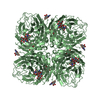
- Structure visualization
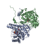
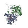
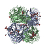
| Structure viewer | Molecule: MolmilJmol/JSmol |
|---|
- Downloads & links
-Download
| PDBx/mmCIF format | 1kbj.cif.gz | 173.6 KB | Display | PDBx/mmCIF format |
|---|---|---|---|---|
| PDB format | pdb1kbj.ent.gz | 136.4 KB | Display | PDB format |
| PDBx/mmJSON format | 1kbj.json.gz | Tree view | PDBx/mmJSON format | |
| Others |  Other downloads Other downloads |
-Validation report
| Arichive directory | https://data.pdbj.org/pub/pdb/validation_reports/kb/1kbjftp://data.pdbj.org/pub/pdb/validation_reports/kb/1kbj | HTTPS FTP |
|---|
-Related structure data
| Related structure data |  1kbiC 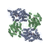 1fcbS S: Starting model for refinement C: citing same article ( |
|---|---|
| Similar structure data |


-Links
 PDBj
PDBj

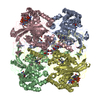
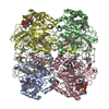
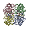
- Assembly
| Deposited unit | 
| ||||||||
|---|---|---|---|---|---|---|---|---|---|
| 1 | 
| ||||||||
| Unit cell |
| ||||||||
| Components on special symmetry positions |
| ||||||||
| Details | The enzyme is a tetramer. |
-Components
| #1: Protein | Mass: 45691.297 Da / Num. of mol.: 2 / Fragment: FMN-BINDING DOMAIN Source method: isolated from a genetically manipulated source Source: (gene. exp.) Production host:  References: UniProt: P00175, L-lactate dehydrogenase (cytochrome) #2: Chemical |   Mass: 456.344 Da / Num. of mol.: 2 / Source method: obtained synthetically / Formula: C17H21N4O9P Mass: 456.344 Da / Num. of mol.: 2 / Source method: obtained synthetically / Formula: C17H21N4O9P#3: Chemical |   Mass: 62.068 Da / Num. of mol.: 2 / Source method: obtained synthetically / Formula: C2H6O2 Mass: 62.068 Da / Num. of mol.: 2 / Source method: obtained synthetically / Formula: C2H6O2#4: Water | ChemComp-HOH / |  Mass: 18.015 Da / Num. of mol.: 370 / Source method: isolated from a natural source / Formula: H2O Mass: 18.015 Da / Num. of mol.: 370 / Source method: isolated from a natural source / Formula: H2O |
|---|
-Experimental details
-Experiment
| Experiment | Method: X-RAY DIFFRACTION / Number of used crystals: 1 |
|---|
- Sample preparation
| Crystal | Density Matthews: 2.9 Å3/Da / Density % sol: 57.58 % | ||||||||||||||||||||||||||||||||||||
|---|---|---|---|---|---|---|---|---|---|---|---|---|---|---|---|---|---|---|---|---|---|---|---|---|---|---|---|---|---|---|---|---|---|---|---|---|---|
| Crystal grow | Temperature: 277 K / Method: vapor diffusion, hanging drop / pH: 5.6 Details: PEG, sodium citrate, ethylene glycol, pH 5.6, VAPOR DIFFUSION, HANGING DROP, temperature 277K | ||||||||||||||||||||||||||||||||||||
| Crystal grow | *PLUS Temperature: 4 ℃ / pH: 7.5 | ||||||||||||||||||||||||||||||||||||
| Components of the solutions | *PLUS
|
-Data collection
| Diffraction | Mean temperature: 100 K |
|---|---|
| Diffraction source | Source: SYNCHROTRON / Site: APS  / Beamline: 19-ID / Wavelength: 1 Å / Beamline: 19-ID / Wavelength: 1 Å |
| Detector | Type: CUSTOM-MADE / Detector: CCD / Date: Apr 9, 1998 |
| Radiation | Protocol: SINGLE WAVELENGTH / Monochromatic (M) / Laue (L): M / Scattering type: x-ray |
| Radiation wavelength | Wavelength: 1 Å / Relative weight: 1 |
| Reflection | Resolution: 2.5→50 Å / Num. all: 37593 / Num. obs: 34160 / % possible obs: 98.8 % / Observed criterion σ(F): -3 / Observed criterion σ(I): -3 / Redundancy: 6.6 % / Rmerge(I) obs: 0.083 / Net I/σ(I): 21.3 |
| Reflection shell | Resolution: 2.5→2.54 Å / Redundancy: 5.8 % / Rmerge(I) obs: 0.345 / Mean I/σ(I) obs: 5.2 / % possible all: 96.2 |
| Reflection | *PLUS Highest resolution: 2.5 Å / Lowest resolution: 50 Å / Num. obs: 36688 / Num. measured all: 240788 |
| Reflection shell | *PLUS Highest resolution: 2.5 Å / % possible obs: 96.2 % |
- Processing
| Software |
| ||||||||||||||||||||||||||||||||||||||||||
|---|---|---|---|---|---|---|---|---|---|---|---|---|---|---|---|---|---|---|---|---|---|---|---|---|---|---|---|---|---|---|---|---|---|---|---|---|---|---|---|---|---|---|---|
| Refinement | Method to determine structure: MOLECULAR REPLACEMENT Starting model: PDB ENTRY 1FCB Resolution: 2.5→40 Å / Isotropic thermal model: isotropic / Cross valid method: THROUGHOUT / σ(F): 0 / σ(I): 0 / Stereochemistry target values: Engh & Huber
| ||||||||||||||||||||||||||||||||||||||||||
| Displacement parameters | Biso mean: 38 Å2 | ||||||||||||||||||||||||||||||||||||||||||
| Refinement step | Cycle: LAST / Resolution: 2.5→40 Å
| ||||||||||||||||||||||||||||||||||||||||||
| Refine LS restraints |
| ||||||||||||||||||||||||||||||||||||||||||
| LS refinement shell |
| ||||||||||||||||||||||||||||||||||||||||||
| Refinement | *PLUS % reflection Rfree: 10 % / Rfactor obs: 0.157 | ||||||||||||||||||||||||||||||||||||||||||
| Solvent computation | *PLUS | ||||||||||||||||||||||||||||||||||||||||||
| Displacement parameters | *PLUS | ||||||||||||||||||||||||||||||||||||||||||
| LS refinement shell | *PLUS Highest resolution: 2.5 Å |
