 Movie
Movie Controller

[English] 日本語
 Yorodumi
- PDB-1m7s: Crystal Structure Analysis of Catalase CatF of Pseudomonas syringae -
+ Open data

- Basic information
| Entry | Database: PDB / ID: 1m7s | ||||||
|---|---|---|---|---|---|---|---|
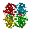
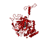
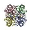
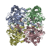
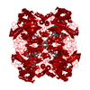
| Title | Crystal Structure Analysis of Catalase CatF of Pseudomonas syringae | ||||||
 Components Components | Catalase | ||||||
 Keywords Keywords | OXIDOREDUCTASE / BETA BARREL / ALPHA HELICAL DOMAIN | ||||||
| Function / homology |  Function and homology information Function and homology informationcatalase / catalase activity / hydrogen peroxide catabolic process / response to hydrogen peroxide / periplasmic space / heme binding / metal ion binding / cytoplasm Similarity search - Function | ||||||
| Biological species |  Pseudomonas syringae (bacteria) Pseudomonas syringae (bacteria) | ||||||
| Method |  X-RAY DIFFRACTION / SYNCHROTRON / MOLECULAR REPLACEMENT / Resolution: 1.8 Å X-RAY DIFFRACTION / SYNCHROTRON / MOLECULAR REPLACEMENT / Resolution: 1.8 Å | ||||||
 Authors Authors | Carpena, X. / Soriano, M. / Klotz, M.G. / Duckworth, H.W. / Donald, L.J. / Melik-Adamyan, W. / Fita, I. / Loewen, P.C. | ||||||
 Citation Citation | Journal: Proteins / Year: 2003 Title: Structure of the Clade 1 catalase, CatF of Pseudomonas syringae, at 1.8 A resolution Authors: Carpena, X. / Soriano, M. / Klotz, M.G. / Duckworth, H.W. / Donald, L.J. / Melik-Adamyan, W. / Fita, I. / Loewen, P.C. | ||||||
| History |
| ||||||
| Remark 999 | SEQUENCE ACCORDING TO THE AUTHOR, THERE WERE ERRORS IN THE ORIGINAL DNA SEQUENCE. THE SEQUENCE ...SEQUENCE ACCORDING TO THE AUTHOR, THERE WERE ERRORS IN THE ORIGINAL DNA SEQUENCE. THE SEQUENCE PRESENTED IN THE ENTRY IS CORRECT. |
- Structure visualization
| Structure viewer | Molecule: MolmilJmol/JSmol |
|---|
- Downloads & links
-Download
| PDBx/mmCIF format | 1m7s.cif.gz | 434.2 KB | Display | PDBx/mmCIF format |
|---|---|---|---|---|
| PDB format | pdb1m7s.ent.gz | 350.1 KB | Display | PDB format |
| PDBx/mmJSON format | 1m7s.json.gz | Tree view | PDBx/mmJSON format | |
| Others |  Other downloads Other downloads |
-Validation report
| Arichive directory | https://data.pdbj.org/pub/pdb/validation_reports/m7/1m7sftp://data.pdbj.org/pub/pdb/validation_reports/m7/1m7s | HTTPS FTP |
|---|
-Related structure data
| Similar structure data |
|---|


-Links
 PDBj
PDBj
- Assembly
| Deposited unit | 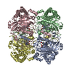
| ||||||||
|---|---|---|---|---|---|---|---|---|---|
| 1 |
| ||||||||
| Unit cell |
|
-Components
| #1: Protein | Mass: 54042.305 Da / Num. of mol.: 4 Source method: isolated from a genetically manipulated source Source: (gene. exp.) Pseudomonas syringae (bacteria) / Gene: CATF / Plasmid: pEC3E56 / Production host: #2: Chemical | ChemComp-HEM /   Mass: 616.487 Da / Num. of mol.: 4 / Source method: obtained synthetically / Formula: C34H32FeN4O4 Mass: 616.487 Da / Num. of mol.: 4 / Source method: obtained synthetically / Formula: C34H32FeN4O4#3: Water | ChemComp-HOH / |  Mass: 18.015 Da / Num. of mol.: 2129 / Source method: isolated from a natural source / Formula: H2O Mass: 18.015 Da / Num. of mol.: 2129 / Source method: isolated from a natural source / Formula: H2O |
|---|
-Experimental details
-Experiment
| Experiment | Method: X-RAY DIFFRACTION / Number of used crystals: 1 |
|---|
- Sample preparation
| Crystal | Density Matthews: 2.3 Å3/Da / Density % sol: 46.44 % | ||||||||||||||||||||||||
|---|---|---|---|---|---|---|---|---|---|---|---|---|---|---|---|---|---|---|---|---|---|---|---|---|---|
| Crystal grow | Temperature: 293 K / Method: vapor diffusion, hanging drop / pH: 6 Details: PEG 4000, sodium cacodylate, pH 6.0, VAPOR DIFFUSION, HANGING DROP, temperature 293K | ||||||||||||||||||||||||
| Crystal grow | *PLUS pH: 6 | ||||||||||||||||||||||||
| Components of the solutions | *PLUS
|
-Data collection
| Diffraction | Mean temperature: 100 K |
|---|---|
| Diffraction source | Source: SYNCHROTRON / Site: ESRF  / Beamline: ID14-2 / Wavelength: 0.933 Å / Beamline: ID14-2 / Wavelength: 0.933 Å |
| Detector | Type: MARRESEARCH / Detector: CCD / Date: Nov 21, 1999 |
| Radiation | Monochromator: DIAMOND CRYSTALS [111] / Protocol: SINGLE WAVELENGTH / Monochromatic (M) / Laue (L): M / Scattering type: x-ray |
| Radiation wavelength | Wavelength: 0.933 Å / Relative weight: 1 |
| Reflection | Resolution: 1.8→30 Å / Num. all: 175279 / Num. obs: 175279 / % possible obs: 97.8 % / Observed criterion σ(I): -3 / Biso Wilson estimate: 22.6 Å2 / Rsym value: 0.084 |
| Reflection shell | Resolution: 1.8→1.87 Å / Rmerge(I) obs: 0.21 / Mean I/σ(I) obs: 5.9 / Num. unique all: 17810 / Rsym value: 0.21 / % possible all: 95.1 |
| Reflection | *PLUS Lowest resolution: 21.9 Å / Rmerge(I) obs: 0.084 |
| Reflection shell | *PLUS % possible obs: 95.1 % / Num. unique obs: 17810 / Rmerge(I) obs: 0.21 |
- Processing
| Software |
| ||||||||||||||||||||
|---|---|---|---|---|---|---|---|---|---|---|---|---|---|---|---|---|---|---|---|---|---|
| Refinement | Method to determine structure: MOLECULAR REPLACEMENT / Resolution: 1.8→21.9 Å / σ(F): 0 / Stereochemistry target values: Engh & Huber
| ||||||||||||||||||||
| Refinement step | Cycle: LAST / Resolution: 1.8→21.9 Å
| ||||||||||||||||||||
| Refinement | *PLUS % reflection Rfree: 5 % / Rfactor Rfree: 0.241 | ||||||||||||||||||||
| Solvent computation | *PLUS | ||||||||||||||||||||
| Displacement parameters | *PLUS | ||||||||||||||||||||
| Refine LS restraints | *PLUS
| ||||||||||||||||||||
| LS refinement shell | *PLUS Rfactor Rfree: 0.294 / Num. reflection Rfree: 900 / Rfactor Rwork: 0.234 / Num. reflection Rwork: 16910 |
