 Movie
Movie Controller


+ Open data

- Basic information
| Entry | Database: PDB / ID: 1k9k | ||||||
|---|---|---|---|---|---|---|---|
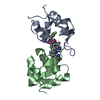
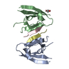
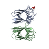
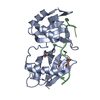
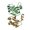
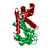
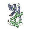
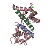
| Title | CRYSTAL STRUCTURE OF CALCIUM BOUND HUMAN S100A6 | ||||||
 Components Components | S100A6 | ||||||
 Keywords Keywords | SIGNALING PROTEIN / S100A6 / CALCYCLIN / CALCIUM REGULATORY PROTEIN / CALCIUM BOUND / CACY | ||||||
| Function / homology |  Function and homology information Function and homology informationmonoatomic ion transmembrane transporter activity / S100 protein binding / tropomyosin binding / ruffle / axonogenesis / positive regulation of fibroblast proliferation / cytoplasmic side of plasma membrane / calcium-dependent protein binding / nuclear envelope / extracellular matrix ...monoatomic ion transmembrane transporter activity / S100 protein binding / tropomyosin binding / ruffle / axonogenesis / positive regulation of fibroblast proliferation / cytoplasmic side of plasma membrane / calcium-dependent protein binding / nuclear envelope / extracellular matrix / calcium ion binding / perinuclear region of cytoplasm / signal transduction / protein homodimerization activity / extracellular exosome / extracellular region / zinc ion binding / nucleus / plasma membrane / cytoplasm / cytosol Similarity search - Function | ||||||
| Biological species |  Homo sapiens (human) Homo sapiens (human) | ||||||
| Method |  X-RAY DIFFRACTION / SYNCHROTRON / MOLECULAR REPLACEMENT / Resolution: 1.76 Å X-RAY DIFFRACTION / SYNCHROTRON / MOLECULAR REPLACEMENT / Resolution: 1.76 Å | ||||||
 Authors Authors | Otterbein, L.R. / Dominguez, R. | ||||||
 Citation Citation | Journal: Structure / Year: 2002 Title: Crystal structures of S100A6 in the Ca(2+)-free and Ca(2+)-bound states: the calcium sensor mechanism of S100 proteins revealed at atomic resolution. Authors: Otterbein, L.R. / Kordowska, J. / Witte-Hoffmann, C. / Wang, C.L. / Dominguez, R. | ||||||
| History |
|
- Structure visualization
| Structure viewer | Molecule: MolmilJmol/JSmol |
|---|
- Downloads & links
-Download
| PDBx/mmCIF format | 1k9k.cif.gz | 52.3 KB | Display | PDBx/mmCIF format |
|---|---|---|---|---|
| PDB format | pdb1k9k.ent.gz | 36.5 KB | Display | PDB format |
| PDBx/mmJSON format | 1k9k.json.gz | Tree view | PDBx/mmJSON format | |
| Others |  Other downloads Other downloads |
-Validation report
| Arichive directory | https://data.pdbj.org/pub/pdb/validation_reports/k9/1k9kftp://data.pdbj.org/pub/pdb/validation_reports/k9/1k9k | HTTPS FTP |
|---|
-Related structure data
| Related structure data | 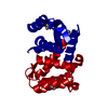 1k8uC  1k96C  1k9pC  1psrS C: citing same article ( S: Starting model for refinement |
|---|---|
| Similar structure data |


-Links
 PDBj
PDBj


- Assembly
| Deposited unit | 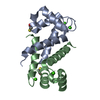
| ||||||||
|---|---|---|---|---|---|---|---|---|---|
| 1 |
| ||||||||
| 2 | 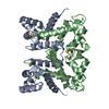
| ||||||||
| Unit cell |
|
-Components
| #1: Protein | Mass: 10193.729 Da / Num. of mol.: 2 Source method: isolated from a genetically manipulated source Source: (gene. exp.) Homo sapiens (human) / Gene: S100A6 / Plasmid: pAED4 / Production host:  #2: Chemical | ChemComp-CA /   Mass: 40.078 Da / Num. of mol.: 4 / Source method: obtained synthetically / Formula: Ca Mass: 40.078 Da / Num. of mol.: 4 / Source method: obtained synthetically / Formula: Ca#3: Chemical |   Mass: 78.133 Da / Num. of mol.: 2 / Source method: obtained synthetically / Formula: C2H6OS Mass: 78.133 Da / Num. of mol.: 2 / Source method: obtained synthetically / Formula: C2H6OS#4: Water | ChemComp-HOH / |  Mass: 18.015 Da / Num. of mol.: 151 / Source method: isolated from a natural source / Formula: H2O Mass: 18.015 Da / Num. of mol.: 151 / Source method: isolated from a natural source / Formula: H2O |
|---|
-Experimental details
-Experiment
| Experiment | Method: X-RAY DIFFRACTION / Number of used crystals: 1 |
|---|
- Sample preparation
| Crystal | Density Matthews: 2.1 Å3/Da / Density % sol: 42 % | |||||||||||||||||||||||||||||||||||||||||||||||||||||||||||||||
|---|---|---|---|---|---|---|---|---|---|---|---|---|---|---|---|---|---|---|---|---|---|---|---|---|---|---|---|---|---|---|---|---|---|---|---|---|---|---|---|---|---|---|---|---|---|---|---|---|---|---|---|---|---|---|---|---|---|---|---|---|---|---|---|---|
| Crystal grow | Temperature: 293 K / Method: vapor diffusion, hanging drop / pH: 7.8 Details: 20% PEG 5000 MME, 30 mM TRIS-HCL, 8% GLYCEROL, pH 7.8, VAPOR DIFFUSION, HANGING DROP, temperature 293K | |||||||||||||||||||||||||||||||||||||||||||||||||||||||||||||||
| Crystal grow | *PLUS Temperature: 20 ℃ / pH: 6.5 | |||||||||||||||||||||||||||||||||||||||||||||||||||||||||||||||
| Components of the solutions | *PLUS
|
-Data collection
| Diffraction | Mean temperature: 100 K |
|---|---|
| Diffraction source | Source: SYNCHROTRON / Site: APS  / Beamline: 14-BM-D / Wavelength: 1 Å / Beamline: 14-BM-D / Wavelength: 1 Å |
| Detector | Type: ADSC QUANTUM 4 / Detector: CCD / Date: Dec 7, 2000 / Details: BENT CYLINDRICAL SI- MIRROR (RH COATING) |
| Radiation | Monochromator: SI(III) DOUBLE CRYSTAL MONOCHROMATOR, L1=1 / Protocol: SINGLE WAVELENGTH / Monochromatic (M) / Laue (L): M / Scattering type: x-ray |
| Radiation wavelength | Wavelength: 1 Å / Relative weight: 1 |
| Reflection | Resolution: 1.75→19.2 Å / Num. all: 17303 / Num. obs: 16853 / % possible obs: 97.4 % / Observed criterion σ(I): 0 / Redundancy: 4.4 % / Biso Wilson estimate: 19.27 Å2 / Rmerge(I) obs: 0.062 / Net I/σ(I): 9.9 |
| Reflection shell | Resolution: 1.75→1.89 Å / Redundancy: 3.5 % / Rmerge(I) obs: 0.192 / Num. unique all: 3218 / % possible all: 96.6 |
| Reflection | *PLUS Num. measured all: 74706 / Rmerge(I) obs: 0.062 |
| Reflection shell | *PLUS Rmerge(I) obs: 0.192 |
- Processing
| Software |
| |||||||||||||||||||||||||
|---|---|---|---|---|---|---|---|---|---|---|---|---|---|---|---|---|---|---|---|---|---|---|---|---|---|---|
| Refinement | Method to determine structure: MOLECULAR REPLACEMENT Starting model: PSORIASIN S100A7 (PBD # 1PSR) Resolution: 1.76→19.2 Å / Cross valid method: THROUGHOUT / σ(F): 0 / σ(I): 0
| |||||||||||||||||||||||||
| Refine analyze | Luzzati sigma a obs: 0.11 Å | |||||||||||||||||||||||||
| Refinement step | Cycle: LAST / Resolution: 1.76→19.2 Å
| |||||||||||||||||||||||||
| Refine LS restraints |
| |||||||||||||||||||||||||
| LS refinement shell | Resolution: 1.76→1.9 Å / Rfactor Rfree error: 0.11
| |||||||||||||||||||||||||
| Refinement | *PLUS % reflection Rfree: 5 % / Rfactor obs: 0.185 / Rfactor Rfree: 0.199 / Rfactor Rwork: 0.185 | |||||||||||||||||||||||||
| Solvent computation | *PLUS | |||||||||||||||||||||||||
| Displacement parameters | *PLUS | |||||||||||||||||||||||||
| LS refinement shell | *PLUS Rfactor Rfree: 0.227 / Rfactor Rwork: 0.206 |
