 Movie
Movie Controller

[English] 日本語
 Yorodumi
- PDB-1k8q: CRYSTAL STRUCTURE OF DOG GASTRIC LIPASE IN COMPLEX WITH A PHOSPHO... -
+ Open data

- Basic information
| Entry | Database: PDB / ID: 1k8q | ||||||
|---|---|---|---|---|---|---|---|
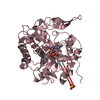
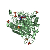
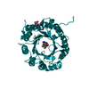
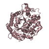
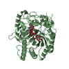
| Title | CRYSTAL STRUCTURE OF DOG GASTRIC LIPASE IN COMPLEX WITH A PHOSPHONATE INHIBITOR | ||||||
 Components Components | Triacylglycerol lipase, gastric | ||||||
 Keywords Keywords | HYDROLASE / APHA BETA HYDROLASE FOLD | ||||||
| Function / homology |  Function and homology information Function and homology informationtriacylglycerol lipase / triacylglycerol lipase activity / lipid catabolic process / lipid metabolic process / extracellular region Similarity search - Function | ||||||
| Biological species |  | ||||||
| Method |  X-RAY DIFFRACTION / SYNCHROTRON / MOLECULAR REPLACEMENT / Resolution: 2.7 Å X-RAY DIFFRACTION / SYNCHROTRON / MOLECULAR REPLACEMENT / Resolution: 2.7 Å | ||||||
 Authors Authors | Roussel, A. / Miled, N. / Berti-Dupuis, L. / Riviere, M. / Spinelli, S. / Berna, P. / Gruber, V. / Verger, R. / Cambillau, C. | ||||||
 Citation Citation | Journal: J.Biol.Chem. / Year: 2002 Title: Crystal structure of the open form of dog gastric lipase in complex with a phosphonate inhibitor. Authors: Roussel, A. / Miled, N. / Berti-Dupuis, L. / Riviere, M. / Spinelli, S. / Berna, P. / Gruber, V. / Verger, R. / Cambillau, C. | ||||||
| History |
|
- Structure visualization
| Structure viewer | Molecule: MolmilJmol/JSmol |
|---|
- Downloads & links
-Download
| PDBx/mmCIF format | 1k8q.cif.gz | 174 KB | Display | PDBx/mmCIF format |
|---|---|---|---|---|
| PDB format | pdb1k8q.ent.gz | 137.6 KB | Display | PDB format |
| PDBx/mmJSON format | 1k8q.json.gz | Tree view | PDBx/mmJSON format | |
| Others |  Other downloads Other downloads |
-Validation report
| Arichive directory | https://data.pdbj.org/pub/pdb/validation_reports/k8/1k8qftp://data.pdbj.org/pub/pdb/validation_reports/k8/1k8q | HTTPS FTP |
|---|
-Related structure data
| Related structure data |  1hlgS S: Starting model for refinement |
|---|---|
| Similar structure data |


-Links
 PDBj
PDBj


- Assembly
| Deposited unit | 
| ||||||||
|---|---|---|---|---|---|---|---|---|---|
| 1 | 
| ||||||||
| 2 | 
| ||||||||
| Unit cell |
|
-Components
| #1: Protein | Mass: 42933.855 Da / Num. of mol.: 2 Source method: isolated from a genetically manipulated source Source: (gene. exp.)  #2: Sugar | ChemComp-NAG /   Type: D-saccharide, beta linking / Mass: 221.208 Da / Num. of mol.: 8 Type: D-saccharide, beta linking / Mass: 221.208 Da / Num. of mol.: 8Source method: isolated from a genetically manipulated source Formula: C8H15NO6 #3: Sugar |   Type: D-saccharide / Mass: 292.369 Da / Num. of mol.: 2 Type: D-saccharide / Mass: 292.369 Da / Num. of mol.: 2Source method: isolated from a genetically manipulated source Formula: C14H28O6 / Comment: detergent*YM #4: Chemical |   Mass: 276.395 Da / Num. of mol.: 2 / Source method: obtained synthetically / Formula: C15H33O2P Mass: 276.395 Da / Num. of mol.: 2 / Source method: obtained synthetically / Formula: C15H33O2P#5: Water | ChemComp-HOH / |  Mass: 18.015 Da / Num. of mol.: 382 / Source method: isolated from a natural source / Formula: H2O Mass: 18.015 Da / Num. of mol.: 382 / Source method: isolated from a natural source / Formula: H2OHas protein modification | Y | |
|---|
-Experimental details
-Experiment
| Experiment | Method: X-RAY DIFFRACTION / Number of used crystals: 1 |
|---|
- Sample preparation
| Crystal | Density Matthews: 2.6 Å3/Da / Density % sol: 52.61 % | ||||||||||||||||||||||||||||||
|---|---|---|---|---|---|---|---|---|---|---|---|---|---|---|---|---|---|---|---|---|---|---|---|---|---|---|---|---|---|---|---|
| Crystal grow | Temperature: 298 K / Method: vapor diffusion, hanging drop / pH: 4.6 Details: 30 % PEGMME 2000, 0.1 M sodium acetate and 0.2 M ammonium sulfate, pH 4.6, VAPOR DIFFUSION, HANGING DROP, temperature 298.0K | ||||||||||||||||||||||||||||||
| Crystal grow | *PLUS | ||||||||||||||||||||||||||||||
| Components of the solutions | *PLUS
|
-Data collection
| Diffraction | Mean temperature: 100 K |
|---|---|
| Diffraction source | Source: SYNCHROTRON / Site: ESRF  / Beamline: BM14 / Wavelength: 0.979 Å / Beamline: BM14 / Wavelength: 0.979 Å |
| Detector | Type: MARRESEARCH / Detector: CCD / Date: Jul 27, 2000 |
| Radiation | Monochromator: mirrors / Protocol: SINGLE WAVELENGTH / Monochromatic (M) / Laue (L): M / Scattering type: x-ray |
| Radiation wavelength | Wavelength: 0.979 Å / Relative weight: 1 |
| Reflection | Resolution: 2.7→40 Å / Num. all: 25123 / Num. obs: 24343 / % possible obs: 97.9 % / Observed criterion σ(F): 1 / Observed criterion σ(I): 1 / Rmerge(I) obs: 0.058 |
| Reflection shell | Resolution: 2.7→2.75 Å / Rmerge(I) obs: 0.215 / % possible all: 88.7 |
| Reflection | *PLUS Lowest resolution: 40 Å / Redundancy: 3 % |
| Reflection shell | *PLUS % possible obs: 97.5 % / Redundancy: 2.2 % / Rmerge(I) obs: 0.212 / Mean I/σ(I) obs: 1.8 |
- Processing
| Software |
| ||||||||||||||||||||
|---|---|---|---|---|---|---|---|---|---|---|---|---|---|---|---|---|---|---|---|---|---|
| Refinement | Method to determine structure: MOLECULAR REPLACEMENT Starting model: PDB ENTRY 1HLG Resolution: 2.7→15 Å / σ(F): 1 / Stereochemistry target values: Engh & Huber
| ||||||||||||||||||||
| Refinement step | Cycle: LAST / Resolution: 2.7→15 Å
| ||||||||||||||||||||
| Refinement | *PLUS Lowest resolution: 15 Å / σ(F): 1 / % reflection Rfree: 5 % / Rfactor obs: 0.192 / Rfactor Rfree: 0.258 | ||||||||||||||||||||
| Solvent computation | *PLUS | ||||||||||||||||||||
| Displacement parameters | *PLUS | ||||||||||||||||||||
| Refine LS restraints | *PLUS
|
