 Movie
Movie Controller


+ Open data

- Basic information
| Entry | Database: PDB / ID: 1k44 | ||||||
|---|---|---|---|---|---|---|---|
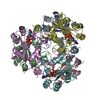
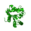
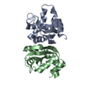
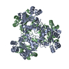
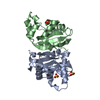
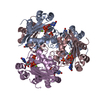
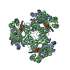
| Title | Mycobacterium tuberculosis Nucleoside Diphosphate Kinase | ||||||
 Components Components | Nucleoside Diphosphate Kinase | ||||||
 Keywords Keywords | TRANSFERASE / Nucleoside Triphosphate / Nucleoside Diphosphate | ||||||
| Function / homology |  Function and homology information Function and homology informationsymbiont-mediated perturbation of host Rab small GTPase signal transduction / purine nucleotide metabolic process / pyrimidine nucleotide metabolic process / symbiont-mediated killing of host cell / nuclease activity / nucleoside-diphosphate kinase / UTP biosynthetic process / CTP biosynthetic process / nucleoside diphosphate kinase activity / GTP biosynthetic process ...symbiont-mediated perturbation of host Rab small GTPase signal transduction / purine nucleotide metabolic process / pyrimidine nucleotide metabolic process / symbiont-mediated killing of host cell / nuclease activity / nucleoside-diphosphate kinase / UTP biosynthetic process / CTP biosynthetic process / nucleoside diphosphate kinase activity / GTP biosynthetic process / phosphoprotein phosphatase activity / Prevention of phagosomal-lysosomal fusion / symbiont-mediated suppression of host innate immune response / extracellular region / ATP binding / metal ion binding / plasma membrane / cytoplasm / cytosol Similarity search - Function | ||||||
| Biological species |   Mycobacterium tuberculosis (bacteria) Mycobacterium tuberculosis (bacteria) | ||||||
| Method |  X-RAY DIFFRACTION / MOLECULAR REPLACEMENT / Resolution: 2.6 Å X-RAY DIFFRACTION / MOLECULAR REPLACEMENT / Resolution: 2.6 Å | ||||||
 Authors Authors | Chen, Y. / Morera, S. / Lascu, I. / Janin, J. | ||||||
 Citation Citation | Journal: Proteins / Year: 2002 Title: X-ray structure of Mycobacterium tuberculosis nucleoside diphosphate kinase Authors: Chen, Y. / Morera, S. / Mocan, J. / Lascu, I. / Janin, J. #1: Journal: Structure / Year: 1995Title: Nucleoside Diphosphate Kinase B Complexed with GDP at 2 A Resolution Authors: Morera, S. / Lacombe, M.L. / Xu, Y. / LeBras, G. / Janin, J. | ||||||
| History |
|
- Structure visualization
| Structure viewer | Molecule: MolmilJmol/JSmol |
|---|
- Downloads & links
-Download
| PDBx/mmCIF format | 1k44.cif.gz | 155.8 KB | Display | PDBx/mmCIF format |
|---|---|---|---|---|
| PDB format | pdb1k44.ent.gz | 126.3 KB | Display | PDB format |
| PDBx/mmJSON format | 1k44.json.gz | Tree view | PDBx/mmJSON format | |
| Others |  Other downloads Other downloads |
-Validation report
| Arichive directory | https://data.pdbj.org/pub/pdb/validation_reports/k4/1k44ftp://data.pdbj.org/pub/pdb/validation_reports/k4/1k44 | HTTPS FTP |
|---|
-Related structure data
| Related structure data | |
|---|---|
| Similar structure data |


-Links
 PDBj
PDBj

- Assembly
| Deposited unit | 
| ||||||||
|---|---|---|---|---|---|---|---|---|---|
| 1 |
| ||||||||
| Unit cell |
|
-Components
| #1: Protein | Mass: 14522.470 Da / Num. of mol.: 6 Source method: isolated from a genetically manipulated source Source: (gene. exp.) Mycobacterium tuberculosis (bacteria) / Production host: References: UniProt: P84284, UniProt: P9WJH7*PLUS, nucleoside-diphosphate kinase #2: Water | ChemComp-HOH / |  Mass: 18.015 Da / Num. of mol.: 107 / Source method: isolated from a natural source / Formula: H2O Mass: 18.015 Da / Num. of mol.: 107 / Source method: isolated from a natural source / Formula: H2O |
|---|
-Experimental details
-Experiment
| Experiment | Method: X-RAY DIFFRACTION / Number of used crystals: 1 |
|---|
- Sample preparation
| Crystal | Density Matthews: 2.78 Å3/Da / Density % sol: 55.82 % | |||||||||||||||||||||||||||||||||||||||||||||||||
|---|---|---|---|---|---|---|---|---|---|---|---|---|---|---|---|---|---|---|---|---|---|---|---|---|---|---|---|---|---|---|---|---|---|---|---|---|---|---|---|---|---|---|---|---|---|---|---|---|---|---|
| Crystal grow | Temperature: 298 K / Method: vapor diffusion, hanging drop / pH: 6 Details: PEG4000, pH 6, VAPOR DIFFUSION, HANGING DROP, temperature 298K | |||||||||||||||||||||||||||||||||||||||||||||||||
| Crystal grow | *PLUS pH: 7.5 | |||||||||||||||||||||||||||||||||||||||||||||||||
| Components of the solutions | *PLUS
|
-Data collection
| Diffraction | Mean temperature: 298 K |
|---|---|
| Diffraction source | Source: ROTATING ANODE / Type: RIGAKU / Wavelength: 1.54 |
| Detector | Type: MARRESEARCH / Detector: IMAGE PLATE / Date: Sep 15, 2001 |
| Radiation | Monochromator: YALE MIRRORS / Protocol: SINGLE WAVELENGTH / Monochromatic (M) / Laue (L): M / Scattering type: x-ray |
| Radiation wavelength | Wavelength: 1.54 Å / Relative weight: 1 |
| Reflection | Resolution: 2.6→20 Å / Num. obs: 30399 / % possible obs: 99.7 % / Observed criterion σ(F): 0 / Observed criterion σ(I): 0 / Redundancy: 5 % / Biso Wilson estimate: 28 Å2 / Rsym value: 0.137 / Net I/σ(I): 11.7 |
| Reflection shell | Resolution: 2.6→2.7 Å / Redundancy: 5 % / Mean I/σ(I) obs: 2.5 / Num. unique all: 2978 / Rsym value: 0.46 / % possible all: 97.5 |
| Reflection | *PLUS Lowest resolution: 20 Å / % possible obs: 99.5 % / Redundancy: 5.7 % |
- Processing
| Software |
| ||||||||||||||||||||
|---|---|---|---|---|---|---|---|---|---|---|---|---|---|---|---|---|---|---|---|---|---|
| Refinement | Method to determine structure: MOLECULAR REPLACEMENT / Resolution: 2.6→20 Å / σ(F): 0 / Stereochemistry target values: Engh & Huber
| ||||||||||||||||||||
| Refine analyze |
| ||||||||||||||||||||
| Refinement step | Cycle: LAST / Resolution: 2.6→20 Å
| ||||||||||||||||||||
| Refine LS restraints |
| ||||||||||||||||||||
| Refinement | *PLUS Lowest resolution: 20 Å / Rfactor obs: 0.205 / Rfactor Rfree: 0.246 / Rfactor Rwork: 0.205 | ||||||||||||||||||||
| Solvent computation | *PLUS | ||||||||||||||||||||
| Displacement parameters | *PLUS | ||||||||||||||||||||
| Refine LS restraints | *PLUS
|
