 Movie
Movie Controller

[English] 日本語
 Yorodumi
- PDB-1wkj: Crystal Structure of Nucleoside Diphosphate Kinase from Thermus t... -
+ Open data

- Basic information
| Entry | Database: PDB / ID: 1wkj | ||||||
|---|---|---|---|---|---|---|---|
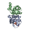
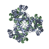
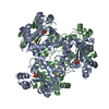
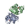
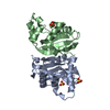
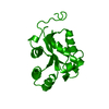
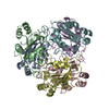
| Title | Crystal Structure of Nucleoside Diphosphate Kinase from Thermus thermophilus HB8 | ||||||
 Components Components | nucleoside diphosphate kinase | ||||||
 Keywords Keywords | TRANSFERASE / nucleoside diphosphate kinase / Thermus thermophilus HB8 / kinase / RIKEN Structural Genomics/Proteomics Initiative / RSGI / Structural Genomics | ||||||
| Function / homology |  Function and homology information Function and homology informationnucleoside-diphosphate kinase / UTP biosynthetic process / CTP biosynthetic process / nucleoside diphosphate kinase activity / GTP biosynthetic process / ATP binding / metal ion binding / cytoplasm Similarity search - Function | ||||||
| Biological species |   Thermus thermophilus (bacteria) Thermus thermophilus (bacteria) | ||||||
| Method |  X-RAY DIFFRACTION / SYNCHROTRON / MOLECULAR REPLACEMENT / Resolution: 2 Å X-RAY DIFFRACTION / SYNCHROTRON / MOLECULAR REPLACEMENT / Resolution: 2 Å | ||||||
 Authors Authors | Takeishi, S. / Nakagawa, N. / Masui, R. / Kuramitsu, S. / RIKEN Structural Genomics/Proteomics Initiative (RSGI) | ||||||
 Citation Citation | Journal: To be Published Title: Crystal Structure of Nucleoside Diphosphate Kinase from Thermus thermophilus HB8 Authors: Takeishi, S. / Nakagawa, N. / Masui, R. / Kuramitsu, S. | ||||||
| History |
|
- Structure visualization
| Structure viewer | Molecule: MolmilJmol/JSmol |
|---|
- Downloads & links
-Download
| PDBx/mmCIF format | 1wkj.cif.gz | 67.6 KB | Display | PDBx/mmCIF format |
|---|---|---|---|---|
| PDB format | pdb1wkj.ent.gz | 50.9 KB | Display | PDB format |
| PDBx/mmJSON format | 1wkj.json.gz | Tree view | PDBx/mmJSON format | |
| Others |  Other downloads Other downloads |
-Validation report
| Arichive directory | https://data.pdbj.org/pub/pdb/validation_reports/wk/1wkjftp://data.pdbj.org/pub/pdb/validation_reports/wk/1wkj | HTTPS FTP |
|---|
-Related structure data
| Related structure data |  1wkkC  1wklC 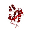 1hhqS C: citing same article ( S: Starting model for refinement |
|---|---|
| Similar structure data | |
| Other databases |
-Links
 PDBj
PDBj- Assembly
| Deposited unit | 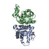
| ||||||||
|---|---|---|---|---|---|---|---|---|---|
| 1 | 
| ||||||||
| Unit cell |
| ||||||||
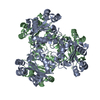
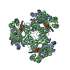
| Details | The biological assembly is a hexamer generated from the dimer in the asymmetric unit by the operations: -y+1, x-y+1, z and -x+y, -x+1, z. |
-Components
| #1: Protein | Mass: 15366.903 Da / Num. of mol.: 2 Source method: isolated from a genetically manipulated source Source: (gene. exp.) Thermus thermophilus (bacteria) / Gene: ndk / Plasmid: pET11a / Species (production host): Escherichia coli / Production host: #2: Water | ChemComp-HOH / |  Mass: 18.015 Da / Num. of mol.: 180 / Source method: isolated from a natural source / Formula: H2O Mass: 18.015 Da / Num. of mol.: 180 / Source method: isolated from a natural source / Formula: H2O |
|---|
-Experimental details
-Experiment
| Experiment | Method: X-RAY DIFFRACTION / Number of used crystals: 1 |
|---|
- Sample preparation
| Crystal | Density Matthews: 3.8 Å3/Da / Density % sol: 67 % |
|---|---|
| Crystal grow | Temperature: 293 K / Method: vapor diffusion, hanging drop / pH: 5.2 Details: 10% PEG4000, 0.09M Ammonium Acetate, 15% Glycerol, 0.1M Sodium Citrate, pH 5.2, VAPOR DIFFUSION, HANGING DROP, temperature 293K |
-Data collection
| Diffraction | Mean temperature: 100 K |
|---|---|
| Diffraction source | Source: SYNCHROTRON / Site: SPring-8  / Beamline: BL45XU / Wavelength: 0.98 Å / Beamline: BL45XU / Wavelength: 0.98 Å |
| Detector | Type: RIGAKU JUPITER 210 / Detector: CCD / Date: Apr 11, 2003 |
| Radiation | Protocol: SINGLE WAVELENGTH / Monochromatic (M) / Laue (L): M / Scattering type: x-ray |
| Radiation wavelength | Wavelength: 0.98 Å / Relative weight: 1 |
| Reflection | Resolution: 2→50 Å / Num. obs: 387285 / % possible obs: 99.4 % / Observed criterion σ(I): -3 / Redundancy: 12 % / Biso Wilson estimate: 27.8 Å2 / Rmerge(I) obs: 0.083 / Net I/σ(I): 39 |
| Reflection shell | Resolution: 2→2.07 Å / Rmerge(I) obs: 0.274 / Mean I/σ(I) obs: 12.7 / % possible all: 100 |
- Processing
| Software |
| |||||||||||||||||||||||||
|---|---|---|---|---|---|---|---|---|---|---|---|---|---|---|---|---|---|---|---|---|---|---|---|---|---|---|
| Refinement | Method to determine structure: MOLECULAR REPLACEMENT Starting model: PDB ENTRY 1HHQ Resolution: 2→50 Å / Isotropic thermal model: Restrained / Cross valid method: THROUGHOUT / σ(F): 0
| |||||||||||||||||||||||||
| Displacement parameters | Biso mean: 29.3 Å2
| |||||||||||||||||||||||||
| Refine analyze |
| |||||||||||||||||||||||||
| Refinement step | Cycle: LAST / Resolution: 2→50 Å
| |||||||||||||||||||||||||
| Refine LS restraints |
|
