 Movie
Movie Controller


+ Open data

- Basic information
| Entry | Database: PDB / ID: 1k3a | ||||||
|---|---|---|---|---|---|---|---|
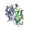
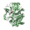
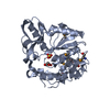
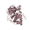
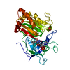
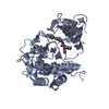
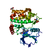
| Title | Structure of the Insulin-like Growth Factor 1 Receptor Kinase | ||||||
 Components Components |
| ||||||
 Keywords Keywords | TRANSFERASE / protein kinase / tyrosine kinase / tyrosine phosphorylation / protein-substrate complex | ||||||
| Function / homology |  Function and homology information Function and homology informationinsulin-like growth factor receptor activity / protein kinase complex / insulin-like growth factor binding / Signaling by Type 1 Insulin-like Growth Factor 1 Receptor (IGF1R) / IRS-related events triggered by IGF1R / positive regulation of fatty acid beta-oxidation / protein transporter activity / positive regulation of glucose metabolic process / transcytosis / IRS-mediated signalling ...insulin-like growth factor receptor activity / protein kinase complex / insulin-like growth factor binding / Signaling by Type 1 Insulin-like Growth Factor 1 Receptor (IGF1R) / IRS-related events triggered by IGF1R / positive regulation of fatty acid beta-oxidation / protein transporter activity / positive regulation of glucose metabolic process / transcytosis / IRS-mediated signalling / insulin receptor complex / insulin-like growth factor I binding / transmembrane receptor protein tyrosine kinase adaptor activity / insulin receptor activity / Activated NTRK3 signals through PI3K / positive regulation of protein-containing complex disassembly / Signaling by Leptin / alphav-beta3 integrin-IGF-1-IGF1R complex / cellular response to fatty acid / peptidyl-tyrosine autophosphorylation / Signaling by LTK / dendritic spine maintenance / PI3K/AKT activation / regulation of JNK cascade / insulin binding / Signaling by ALK / IRS activation / amyloid-beta clearance / Respiratory syncytial virus (RSV) attachment and entry / intracellular membrane-bounded organelle / insulin receptor substrate binding / PI3K Cascade / positive regulation of glycogen biosynthetic process / positive regulation of insulin receptor signaling pathway / SOS-mediated signalling / Signal attenuation / Growth hormone receptor signaling / negative regulation of insulin secretion / SHC-related events triggered by IGF1R / phosphatidylinositol 3-kinase binding / insulin-like growth factor receptor binding / negative regulation of MAPK cascade / phosphotyrosine residue binding / insulin-like growth factor receptor signaling pathway / negative regulation of insulin receptor signaling pathway / signaling adaptor activity / Interleukin-7 signaling / SH2 domain binding / positive regulation of D-glucose import across plasma membrane / protein kinase C binding / insulin receptor binding / cellular response to glucose stimulus / phosphatidylinositol 3-kinase/protein kinase B signal transduction / receptor protein-tyrosine kinase / response to insulin / caveola / cellular response to amyloid-beta / cellular response to insulin stimulus / Constitutive Signaling by Aberrant PI3K in Cancer / cytokine-mediated signaling pathway / insulin receptor signaling pathway / Signaling by ALK fusions and activated point mutants / PIP3 activates AKT signaling / protein autophosphorylation / glucose homeostasis / positive regulation of cold-induced thermogenesis / PI5P, PP2A and IER3 Regulate PI3K/AKT Signaling / signaling receptor complex adaptor activity / RAF/MAP kinase cascade / protein tyrosine kinase activity / positive regulation of MAPK cascade / positive regulation of phosphatidylinositol 3-kinase/protein kinase B signal transduction / cilium / signaling receptor complex / Extra-nuclear estrogen signaling / immune response / positive regulation of cell migration / axon / positive regulation of cell population proliferation / negative regulation of apoptotic process / nucleolus / signal transduction / nucleoplasm / ATP binding / membrane / identical protein binding / nucleus / plasma membrane / cytosol / cytoplasm Similarity search - Function | ||||||
| Biological species |  Homo sapiens (human) Homo sapiens (human) | ||||||
| Method |  X-RAY DIFFRACTION / SYNCHROTRON / MOLECULAR REPLACEMENT / Resolution: 2.1 Å X-RAY DIFFRACTION / SYNCHROTRON / MOLECULAR REPLACEMENT / Resolution: 2.1 Å | ||||||
 Authors Authors | Favelyukis, S. / Till, J.H. / Hubbard, S.R. / Miller, W.T. | ||||||
 Citation Citation | Journal: Nat.Struct.Biol. / Year: 2001 Title: Structure and autoregulation of the insulin-like growth factor 1 receptor kinase. Authors: Favelyukis, S. / Till, J.H. / Hubbard, S.R. / Miller, W.T. | ||||||
| History |
| ||||||
| Remark 999 | SEQUENCE THE RESIDUE CORRESPONDS TO RESIDUE FROM A SYNTHETIC PEPTIDE. IT WAS ADDED TO MODIFY THE ...SEQUENCE THE RESIDUE CORRESPONDS TO RESIDUE FROM A SYNTHETIC PEPTIDE. IT WAS ADDED TO MODIFY THE SEQUENCE TO BE SUITABLE FOR ACTIVITY ASSAYS |
- Structure visualization
| Structure viewer | Molecule: MolmilJmol/JSmol |
|---|
- Downloads & links
-Download
| PDBx/mmCIF format | 1k3a.cif.gz | 78 KB | Display | PDBx/mmCIF format |
|---|---|---|---|---|
| PDB format | pdb1k3a.ent.gz | 56.7 KB | Display | PDB format |
| PDBx/mmJSON format | 1k3a.json.gz | Tree view | PDBx/mmJSON format | |
| Others |  Other downloads Other downloads |
-Validation report
| Arichive directory | https://data.pdbj.org/pub/pdb/validation_reports/k3/1k3aftp://data.pdbj.org/pub/pdb/validation_reports/k3/1k3a | HTTPS FTP |
|---|
-Related structure data
| Related structure data |  1ir3S S: Starting model for refinement |
|---|---|
| Similar structure data |
-Links
 PDBj
PDBj


- Assembly
| Deposited unit | 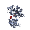
| ||||||||
|---|---|---|---|---|---|---|---|---|---|
| 1 |
| ||||||||
| Unit cell |
| ||||||||
| Details | tetramer of 2 alpha and 2 beta chains linked by disulfide bonds. |
-Components
| #1: Protein | Mass: 34410.199 Da / Num. of mol.: 1 / Fragment: beta chain, kinase domain (residues 988-1286) Source method: isolated from a genetically manipulated source Source: (gene. exp.) Homo sapiens (human) / Plasmid: pFast-Bac / Production host:   Spodoptera frugiperda (fall armyworm) / Strain (production host): Sf9 / References: UniProt: P08069, EC: 2.7.1.112 Spodoptera frugiperda (fall armyworm) / Strain (production host): Sf9 / References: UniProt: P08069, EC: 2.7.1.112 |
|---|---|
| #2: Protein/peptide | Mass: 1598.818 Da / Num. of mol.: 1 / Source method: obtained synthetically / Details: Chemically synthesized / References: UniProt: P35568 |
| #3: Chemical | ChemComp-ACP /   Mass: 505.208 Da / Num. of mol.: 1 / Source method: obtained synthetically / Formula: C11H18N5O12P3 / Comment: AMP-PCP, energy-carrying molecule analogue*YM Mass: 505.208 Da / Num. of mol.: 1 / Source method: obtained synthetically / Formula: C11H18N5O12P3 / Comment: AMP-PCP, energy-carrying molecule analogue*YM |
| #4: Water | ChemComp-HOH /  Mass: 18.015 Da / Num. of mol.: 158 / Source method: isolated from a natural source / Formula: H2O Mass: 18.015 Da / Num. of mol.: 158 / Source method: isolated from a natural source / Formula: H2O |
| Has protein modification | Y |
-Experimental details
-Experiment
| Experiment | Method: X-RAY DIFFRACTION / Number of used crystals: 1 |
|---|
- Sample preparation
| Crystal | Density Matthews: 2.9 Å3/Da / Density % sol: 57.55 % | ||||||||||||||||||||||||||||||||||||||||||||||||||||||||
|---|---|---|---|---|---|---|---|---|---|---|---|---|---|---|---|---|---|---|---|---|---|---|---|---|---|---|---|---|---|---|---|---|---|---|---|---|---|---|---|---|---|---|---|---|---|---|---|---|---|---|---|---|---|---|---|---|---|
| Crystal grow | Temperature: 277 K / Method: vapor diffusion, hanging drop / pH: 7.5 Details: PEG 8000, sodium chloride, HEPES, pH 7.5, VAPOR DIFFUSION, HANGING DROP, temperature 277K | ||||||||||||||||||||||||||||||||||||||||||||||||||||||||
| Crystal grow | *PLUS Temperature: 4 ℃ | ||||||||||||||||||||||||||||||||||||||||||||||||||||||||
| Components of the solutions | *PLUS
|
-Data collection
| Diffraction | Mean temperature: 100 K |
|---|---|
| Diffraction source | Source: SYNCHROTRON / Site: NSLS  / Beamline: X4A / Wavelength: 0.9725 Å / Beamline: X4A / Wavelength: 0.9725 Å |
| Detector | Type: ADSC QUANTUM 4 / Detector: CCD / Date: May 22, 2001 |
| Radiation | Monochromator: silicon crystal / Protocol: SINGLE WAVELENGTH / Monochromatic (M) / Laue (L): M / Scattering type: x-ray |
| Radiation wavelength | Wavelength: 0.9725 Å / Relative weight: 1 |
| Reflection | Resolution: 2.1→28.07 Å / Num. all: 24941 / Num. obs: 24941 / % possible obs: 99.9 % / Observed criterion σ(F): 0 / Observed criterion σ(I): 0 / Biso Wilson estimate: 22.3 Å2 / Rsym value: 0.048 |
| Reflection shell | Resolution: 2.1→2.18 Å / Rsym value: 0.254 / % possible all: 100 |
| Reflection | *PLUS Lowest resolution: 25 Å / Num. measured all: 279673 / Rmerge(I) obs: 0.048 |
| Reflection shell | *PLUS % possible obs: 100 % / Rmerge(I) obs: 0.254 |
- Processing
| Software |
| ||||||||||||||||||||||||||||||||||||||||
|---|---|---|---|---|---|---|---|---|---|---|---|---|---|---|---|---|---|---|---|---|---|---|---|---|---|---|---|---|---|---|---|---|---|---|---|---|---|---|---|---|---|
| Refinement | Method to determine structure: MOLECULAR REPLACEMENT Starting model: 1ir3 Resolution: 2.1→28.07 Å / Rfactor Rfree error: 0.005 / Data cutoff high absF: 387254.78 / Data cutoff low absF: 0 / Isotropic thermal model: RESTRAINED / Cross valid method: THROUGHOUT / σ(F): 0 / Stereochemistry target values: Engh & Huber
| ||||||||||||||||||||||||||||||||||||||||
| Solvent computation | Solvent model: FLAT MODEL / Bsol: 46.8196 Å2 / ksol: 0.357892 e/Å3 | ||||||||||||||||||||||||||||||||||||||||
| Displacement parameters | Biso mean: 33.9 Å2
| ||||||||||||||||||||||||||||||||||||||||
| Refine analyze |
| ||||||||||||||||||||||||||||||||||||||||
| Refinement step | Cycle: LAST / Resolution: 2.1→28.07 Å
| ||||||||||||||||||||||||||||||||||||||||
| Refine LS restraints |
| ||||||||||||||||||||||||||||||||||||||||
| LS refinement shell | Resolution: 2.1→2.23 Å / Rfactor Rfree error: 0.014 / Total num. of bins used: 6
| ||||||||||||||||||||||||||||||||||||||||
| Xplor file |
| ||||||||||||||||||||||||||||||||||||||||
| Software | *PLUS Name: CNS / Version: 1 / Classification: refinement | ||||||||||||||||||||||||||||||||||||||||
| Refinement | *PLUS Lowest resolution: 25 Å / σ(F): 0 / % reflection Rfree: 9.8 % / Rfactor Rfree: 0.248 | ||||||||||||||||||||||||||||||||||||||||
| Solvent computation | *PLUS | ||||||||||||||||||||||||||||||||||||||||
| Displacement parameters | *PLUS Biso mean: 33.9 Å2 | ||||||||||||||||||||||||||||||||||||||||
| Refine LS restraints | *PLUS
| ||||||||||||||||||||||||||||||||||||||||
| LS refinement shell | *PLUS Rfactor Rfree: 0.275 / % reflection Rfree: 9.9 % / Rfactor Rwork: 0.231 |
