| 登録情報 | データベース: PDB / ID: 1k04
|
|---|
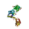
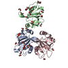
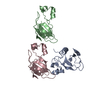
| タイトル | Crystal Structure of the Focal Adhesion Targeting Domain of Focal Adhesion Kinase |
|---|
 要素 要素 | FOCAL ADHESION KINASE 1 |
|---|
 キーワード キーワード | TRANSFERASE / up-down-up-down four helical bundle forming a helix-exchange dimer |
|---|
| 機能・相同性 |  機能・相同性情報 機能・相同性情報
netrin-activated signaling pathway / regulation of substrate adhesion-dependent cell spreading / regulation of endothelial cell migration / regulation of epithelial cell migration / detection of muscle stretch / JUN kinase binding / signal complex assembly / positive regulation of ubiquitin-dependent protein catabolic process / positive regulation of macrophage proliferation / DCC mediated attractive signaling ...netrin-activated signaling pathway / regulation of substrate adhesion-dependent cell spreading / regulation of endothelial cell migration / regulation of epithelial cell migration / detection of muscle stretch / JUN kinase binding / signal complex assembly / positive regulation of ubiquitin-dependent protein catabolic process / positive regulation of macrophage proliferation / DCC mediated attractive signaling / positive regulation of fibroblast migration / Signal regulatory protein family interactions / regulation of GTPase activity / growth hormone receptor signaling pathway / MET activates PTK2 signaling / regulation of focal adhesion assembly / negative regulation of cell-cell adhesion / positive regulation of wound healing / p130Cas linkage to MAPK signaling for integrins / regulation of osteoblast differentiation / Apoptotic cleavage of cellular proteins / establishment of cell polarity / positive regulation of macrophage chemotaxis / regulation of cytoskeleton organization / regulation of cell adhesion mediated by integrin / Fc-gamma receptor signaling pathway involved in phagocytosis / vascular endothelial cell response to oscillatory fluid shear stress / GRB2:SOS provides linkage to MAPK signaling for Integrins / regulation of protein phosphorylation / negative regulation of anoikis / positive regulation of epithelial cell migration / ephrin receptor signaling pathway / Estrogen-dependent nuclear events downstream of ESR-membrane signaling / RHO GTPases Activate WASPs and WAVEs / positive regulation of protein kinase activity / vascular endothelial growth factor receptor signaling pathway / regulation of cell adhesion / heart morphogenesis / positive regulation of epithelial to mesenchymal transition / stress fiber / EPHB-mediated forward signaling / Integrin signaling / protein tyrosine phosphatase activity / NCAM signaling for neurite out-growth / transforming growth factor beta receptor signaling pathway / SH2 domain binding / axon guidance / molecular function activator activity / Turbulent (oscillatory, disturbed) flow shear stress activates signaling by PIEZO1 and integrins in endothelial cells / integrin-mediated signaling pathway / non-membrane spanning protein tyrosine kinase activity / FCGR3A-mediated phagocytosis / non-specific protein-tyrosine kinase / placenta development / cell motility / peptidyl-tyrosine phosphorylation / Regulation of actin dynamics for phagocytic cup formation / VEGFA-VEGFR2 Pathway / epidermal growth factor receptor signaling pathway / integrin binding / cell migration / regulation of cell shape / regulation of cell population proliferation / positive regulation of protein phosphorylation / actin binding / RAF/MAP kinase cascade / cell cortex / protein autophosphorylation / protein tyrosine kinase activity / protein phosphatase binding / angiogenesis / cytoskeleton / Extra-nuclear estrogen signaling / positive regulation of phosphatidylinositol 3-kinase/protein kinase B signal transduction / ciliary basal body / cilium / positive regulation of cell migration / focal adhesion / intracellular membrane-bounded organelle / positive regulation of cell population proliferation / centrosome / negative regulation of apoptotic process / protein kinase binding / perinuclear region of cytoplasm / ATP binding / nucleus / plasma membrane / cytosol類似検索 - 分子機能 Single helix bin / Nucleotidyltransferases domain 2 / Focal adhesion kinase, targeting (FAT) domain / Focal adhesion kinase, targeting (FAT) domain superfamily / Focal adhesion kinase, N-terminal / FAK1/PYK2, FERM domain C-lobe / Focal adhesion targeting region / FERM N-terminal domain / : / FAK1/PYK2, FERM domain C-lobe ...Single helix bin / Nucleotidyltransferases domain 2 / Focal adhesion kinase, targeting (FAT) domain / Focal adhesion kinase, targeting (FAT) domain superfamily / Focal adhesion kinase, N-terminal / FAK1/PYK2, FERM domain C-lobe / Focal adhesion targeting region / FERM N-terminal domain / : / FAK1/PYK2, FERM domain C-lobe / FERM domain signature 2. / FERM central domain / FERM/acyl-CoA-binding protein superfamily / FERM central domain / FERM superfamily, second domain / FERM domain / FERM domain profile. / Band 4.1 domain / Band 4.1 homologues / Single alpha-helices involved in coiled-coils or other helix-helix interfaces / Four Helix Bundle (Hemerythrin (Met), subunit A) / Tyrosine-protein kinase, catalytic domain / Tyrosine kinase, catalytic domain / Tyrosine protein kinases specific active-site signature. / Tyrosine-protein kinase, active site / PH-like domain superfamily / Serine-threonine/tyrosine-protein kinase, catalytic domain / Protein tyrosine and serine/threonine kinase / Ubiquitin-like domain superfamily / Protein kinase, ATP binding site / Protein kinases ATP-binding region signature. / Protein kinase domain profile. / Protein kinase domain / Protein kinase-like domain superfamily / Up-down Bundle / Mainly Alpha類似検索 - ドメイン・相同性 |
|---|
| 生物種 |  Homo sapiens (ヒト) Homo sapiens (ヒト) |
|---|
| 手法 |  X線回折 / シンクロトロン / 分子置換 / 解像度: 1.95 Å X線回折 / シンクロトロン / 分子置換 / 解像度: 1.95 Å |
|---|
 データ登録者 データ登録者 | Arold, S.T. / Hoellerer, M.K. / Noble, M.E.M. |
|---|
 引用 引用 | ジャーナル: Structure / 年: 2002
タイトル: The structural basis of localization and signaling by the focal adhesion targeting domain.
著者: Arold, S.T. / Hoellerer, M.K. / Noble, M.E. |
|---|
| 履歴 | | 登録 | 2001年9月18日 | 登録サイト: RCSB / 処理サイト: RCSB |
|---|
| 改定 1.0 | 2002年1月30日 | Provider: repository / タイプ: Initial release |
|---|
| 改定 1.1 | 2008年4月27日 | Group: Version format compliance |
|---|
| 改定 1.2 | 2011年7月13日 | Group: Derived calculations / Version format compliance |
|---|
| 改定 1.3 | 2024年2月7日 | Group: Data collection / Database references / Derived calculations
カテゴリ: chem_comp_atom / chem_comp_bond ...chem_comp_atom / chem_comp_bond / database_2 / pdbx_struct_special_symmetry / struct_site
Item: _database_2.pdbx_DOI / _database_2.pdbx_database_accession ..._database_2.pdbx_DOI / _database_2.pdbx_database_accession / _struct_site.pdbx_auth_asym_id / _struct_site.pdbx_auth_comp_id / _struct_site.pdbx_auth_seq_id |
|---|
|
|---|
 ムービー
ムービー コントローラー


 データを開く
 基本情報
基本情報 構造の表示
構造の表示 ダウンロードとリンク
 その他のダウンロード


 PDBj
PDBj


 集合体
集合体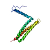


 分子量: 35.453 Da / 分子数: 1 / 由来タイプ: 合成 / 式: Cl
 分子量: 18.015 Da / 分子数: 97 / 由来タイプ: 天然 / 式: H2O
 試料調製
試料調製 / ビームライン: ID14-3 / 波長: 0.931 Å
 解析
解析