 Movie
Movie Controller


+ Open data

- Basic information
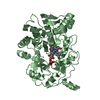
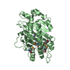
| Entry | Database: PDB / ID: 1k02 | ||||||
|---|---|---|---|---|---|---|---|
| Title | Crystal Structure of Old Yellow Enzyme Mutant Gln114Asn | ||||||
 Components Components | NADPH DEHYDROGENASE 1 | ||||||
 Keywords Keywords | OXIDOREDUCTASE / beta-alpha barrels | ||||||
| Function / homology |  Function and homology information Function and homology informationNADPH dehydrogenase / NADPH dehydrogenase activity / FMN binding / apoptotic process Similarity search - Function | ||||||
| Biological species |  | ||||||
| Method |  X-RAY DIFFRACTION / MOLECULAR REPLACEMENT / Resolution: 2.7 Å X-RAY DIFFRACTION / MOLECULAR REPLACEMENT / Resolution: 2.7 Å | ||||||
 Authors Authors | Brown, B.J. / Hyun, J. / Duvvuri, S.D. / Karplus, P.A. / Massey, V. | ||||||
 Citation Citation | Journal: J.Biol.Chem. / Year: 2002 Title: The role of glutamine 114 in old yellow enzyme Authors: Brown, B.J. / Hyun, J. / Duvvuri, S.D. / Karplus, P.A. / Massey, V. | ||||||
| History |
|
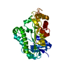
- Structure visualization
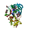
| Structure viewer | Molecule: MolmilJmol/JSmol |
|---|
- Downloads & links
-Download
| PDBx/mmCIF format | 1k02.cif.gz | 102.3 KB | Display | PDBx/mmCIF format |
|---|---|---|---|---|
| PDB format | pdb1k02.ent.gz | 78.7 KB | Display | PDB format |
| PDBx/mmJSON format | 1k02.json.gz | Tree view | PDBx/mmJSON format | |
| Others |  Other downloads Other downloads |
-Validation report
| Arichive directory | https://data.pdbj.org/pub/pdb/validation_reports/k0/1k02ftp://data.pdbj.org/pub/pdb/validation_reports/k0/1k02 | HTTPS FTP |
|---|
-Related structure data


-Links
 PDBj
PDBj- Assembly
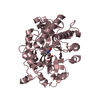
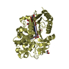
| Deposited unit | 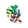
| ||||||||
|---|---|---|---|---|---|---|---|---|---|
| 1 |
| ||||||||
| Unit cell |
|
-Components
| #1: Protein | Mass: 44926.418 Da / Num. of mol.: 1 / Mutation: Q114N Source method: isolated from a genetically manipulated source Source: (gene. exp.) Production host:  |
|---|---|
| #2: Chemical | ChemComp-MG /   Mass: 24.305 Da / Num. of mol.: 1 / Source method: obtained synthetically / Formula: Mg Mass: 24.305 Da / Num. of mol.: 1 / Source method: obtained synthetically / Formula: Mg |
| #3: Chemical | ChemComp-FMN /   Mass: 456.344 Da / Num. of mol.: 1 / Source method: obtained synthetically / Formula: C17H21N4O9P Mass: 456.344 Da / Num. of mol.: 1 / Source method: obtained synthetically / Formula: C17H21N4O9P |
| #4: Water | ChemComp-HOH /  Mass: 18.015 Da / Num. of mol.: 449 / Source method: isolated from a natural source / Formula: H2O Mass: 18.015 Da / Num. of mol.: 449 / Source method: isolated from a natural source / Formula: H2O |
-Experimental details
-Experiment
| Experiment | Method: X-RAY DIFFRACTION / Number of used crystals: 1 |
|---|
- Sample preparation
| Crystal | Density Matthews: 2.42 Å3/Da / Density % sol: 49.25 % | ||||||||||||||||||||||||||||||||||||||||||||||||
|---|---|---|---|---|---|---|---|---|---|---|---|---|---|---|---|---|---|---|---|---|---|---|---|---|---|---|---|---|---|---|---|---|---|---|---|---|---|---|---|---|---|---|---|---|---|---|---|---|---|
| Crystal grow | Temperature: 277 K / Method: vapor diffusion, hanging drop / pH: 8.3 Details: PEG-400, HEPES, Magnesium Chloride, pH 8.3, VAPOR DIFFUSION, HANGING DROP, temperature 277K | ||||||||||||||||||||||||||||||||||||||||||||||||
| Crystal grow | *PLUS Temperature: 4 ℃ / pH: 8 / Details: Fox, K.M., (1993) J. Mol. Biol., 234, 502. | ||||||||||||||||||||||||||||||||||||||||||||||||
| Components of the solutions | *PLUS
|
-Data collection
| Diffraction | Mean temperature: 298 K |
|---|---|
| Diffraction source | Source: ROTATING ANODE / Type: RIGAKU / Wavelength: 1.5418 Å |
| Detector | Type: RIGAKU RAXIS IV / Detector: IMAGE PLATE / Date: Oct 31, 2000 |
| Radiation | Monochromator: Ni MIRROR + Ni FILTER / Protocol: SINGLE WAVELENGTH / Monochromatic (M) / Laue (L): M / Scattering type: x-ray |
| Radiation wavelength | Wavelength: 1.5418 Å / Relative weight: 1 |
| Reflection | Resolution: 2.7→50 Å / Num. all: 12522 / Num. obs: 10792 / % possible obs: 85.2 % / Observed criterion σ(F): 2 / Observed criterion σ(I): 2 |
| Reflection shell | Resolution: 2.7→2.8 Å / Num. unique all: 1227 / % possible all: 99.8 |
| Reflection | *PLUS Lowest resolution: 50 Å / % possible obs: 97.4 % / Num. measured all: 56163 / Rmerge(I) obs: 0.128 |
| Reflection shell | *PLUS % possible obs: 99.8 % / Rmerge(I) obs: 0.53 |
- Processing
| Software |
| ||||||||||||||||
|---|---|---|---|---|---|---|---|---|---|---|---|---|---|---|---|---|---|
| Refinement | Method to determine structure: MOLECULAR REPLACEMENT / Resolution: 2.7→500 Å / σ(F): 2 / Stereochemistry target values: EMBL
| ||||||||||||||||
| Refinement step | Cycle: LAST / Resolution: 2.7→500 Å
| ||||||||||||||||
| Refine LS restraints |
| ||||||||||||||||
| Software | *PLUS Name: CNS / Classification: refinement | ||||||||||||||||
| Refinement | *PLUS Highest resolution: 2.7 Å / Lowest resolution: 500 Å / σ(F): 2 | ||||||||||||||||
| Solvent computation | *PLUS | ||||||||||||||||
| Displacement parameters | *PLUS | ||||||||||||||||
| Refine LS restraints | *PLUS Type: c_angle_deg / Dev ideal: 1.3 |
