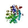
 Movie
Movie Controller


+ Open data

- Basic information
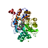
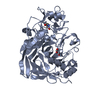
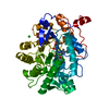
| Entry | Database: PDB / ID: 1bwl | ||||||
|---|---|---|---|---|---|---|---|
| Title | OLD YELLOW ENZYME (OYE1) DOUBLE MUTANT H191N:N194H | ||||||
 Components Components | PROTEIN (NADPH DEHYDROGENASE 1) | ||||||
 Keywords Keywords | OXIDOREDUCTASE / FLAVOENZYME / NADPH OXIDOREDUCTASE / TIM BARREL | ||||||
| Function / homology |  Function and homology information Function and homology informationNADPH dehydrogenase / NADPH dehydrogenase activity / FMN binding / apoptotic process Similarity search - Function | ||||||
| Biological species |  Candida albicans (yeast) Candida albicans (yeast) | ||||||
| Method |  X-RAY DIFFRACTION / OTHER / Resolution: 2.7 Å X-RAY DIFFRACTION / OTHER / Resolution: 2.7 Å | ||||||
 Authors Authors | Brown, B.J. / Deng, Z. / Karplus, P.A. / Massey, V. | ||||||
 Citation Citation | Journal: J.Biol.Chem. / Year: 1998 Title: On the active site of Old Yellow Enzyme. Role of histidine 191 and asparagine 194. Authors: Brown, B.J. / Deng, Z. / Karplus, P.A. / Massey, V. | ||||||
| History |
|
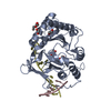
- Structure visualization
| Structure viewer | Molecule: MolmilJmol/JSmol |
|---|
- Downloads & links
-Download
| PDBx/mmCIF format | 1bwl.cif.gz | 93.6 KB | Display | PDBx/mmCIF format |
|---|---|---|---|---|
| PDB format | pdb1bwl.ent.gz | 71.1 KB | Display | PDB format |
| PDBx/mmJSON format | 1bwl.json.gz | Tree view | PDBx/mmJSON format | |
| Others |  Other downloads Other downloads |
-Validation report
| Arichive directory | https://data.pdbj.org/pub/pdb/validation_reports/bw/1bwlftp://data.pdbj.org/pub/pdb/validation_reports/bw/1bwl | HTTPS FTP |
|---|
-Related structure data
| Related structure data | 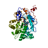 1bwkC  1oyaS S: Starting model for refinement C: citing same article ( |
|---|---|
| Similar structure data |


-Links
 PDBj
PDBj- Assembly
| Deposited unit | 
| ||||||||
|---|---|---|---|---|---|---|---|---|---|
| 1 |
| ||||||||
| Unit cell |
|
-Components
| #1: Protein | Mass: 44898.344 Da / Num. of mol.: 1 / Mutation: H191N:N194H Source method: isolated from a genetically manipulated source Source: (gene. exp.) Candida albicans (yeast) / Gene: OYE1 / Plasmid: PET3B / Gene (production host): OYE1-H191N:N194H / Production host:  |
|---|---|
| #2: Chemical | ChemComp-FMN /   Mass: 456.344 Da / Num. of mol.: 1 / Source method: obtained synthetically / Formula: C17H21N4O9P Mass: 456.344 Da / Num. of mol.: 1 / Source method: obtained synthetically / Formula: C17H21N4O9P |
| #3: Water | ChemComp-HOH /  Mass: 18.015 Da / Num. of mol.: 127 / Source method: isolated from a natural source / Formula: H2O Mass: 18.015 Da / Num. of mol.: 127 / Source method: isolated from a natural source / Formula: H2O |
-Experimental details
-Experiment
| Experiment | Method: X-RAY DIFFRACTION |
|---|
- Sample preparation
| Crystal | Density Matthews: 2.46 Å3/Da / Density % sol: 43 % | ||||||||||||||||||||||||||||||||||||||||||||||||
|---|---|---|---|---|---|---|---|---|---|---|---|---|---|---|---|---|---|---|---|---|---|---|---|---|---|---|---|---|---|---|---|---|---|---|---|---|---|---|---|---|---|---|---|---|---|---|---|---|---|
| Crystal grow | pH: 8.3 / Details: pH 8.3 | ||||||||||||||||||||||||||||||||||||||||||||||||
| Crystal | *PLUS | ||||||||||||||||||||||||||||||||||||||||||||||||
| Crystal grow | *PLUS Temperature: 4 ℃ / pH: 8 / Method: vapor diffusion, hanging drop / Details: Fox, K.M., (1993) J. Mol. Biol., 234, 502. | ||||||||||||||||||||||||||||||||||||||||||||||||
| Components of the solutions | *PLUS
|
-Data collection
| Diffraction | Mean temperature: 293 K |
|---|---|
| Diffraction source | Source: ROTATING ANODE / Type: RIGAKU RU200 / Wavelength: 1.5418 |
| Detector | Type: SDMS / Detector: AREA DETECTOR |
| Radiation | Protocol: SINGLE WAVELENGTH / Monochromatic (M) / Laue (L): M / Scattering type: x-ray |
| Radiation wavelength | Wavelength: 1.5418 Å / Relative weight: 1 |
| Reflection | Resolution: 2.7→25 Å / Num. obs: 11781 / % possible obs: 91.4 % / Redundancy: 3.6 % / Rsym value: 0.117 / Net I/σ(I): 11.1 |
| Reflection shell | Resolution: 2.7→2.8 Å / Mean I/σ(I) obs: 3.3 / Rsym value: 0.303 / % possible all: 82.1 |
| Reflection | *PLUS Num. measured all: 42885 / Rmerge(I) obs: 0.117 |
| Reflection shell | *PLUS % possible obs: 82.1 % / Rmerge(I) obs: 0.303 |
- Processing
| Software |
| ||||||||||||||||||||||||||||||||||||||||||||||||||||||||||||
|---|---|---|---|---|---|---|---|---|---|---|---|---|---|---|---|---|---|---|---|---|---|---|---|---|---|---|---|---|---|---|---|---|---|---|---|---|---|---|---|---|---|---|---|---|---|---|---|---|---|---|---|---|---|---|---|---|---|---|---|---|---|
| Refinement | Method to determine structure: OTHER Starting model: PDB ENTRY 1OYA Resolution: 2.7→8 Å / Data cutoff high absF: 10000000 / Data cutoff low absF: 0.001 / Cross valid method: THROUGHOUT / σ(F): 2
| ||||||||||||||||||||||||||||||||||||||||||||||||||||||||||||
| Displacement parameters | Biso mean: 15.5 Å2 | ||||||||||||||||||||||||||||||||||||||||||||||||||||||||||||
| Refinement step | Cycle: LAST / Resolution: 2.7→8 Å
| ||||||||||||||||||||||||||||||||||||||||||||||||||||||||||||
| Refine LS restraints |
| ||||||||||||||||||||||||||||||||||||||||||||||||||||||||||||
| LS refinement shell | Resolution: 2.7→2.82 Å / Total num. of bins used: 8
| ||||||||||||||||||||||||||||||||||||||||||||||||||||||||||||
| Xplor file |
| ||||||||||||||||||||||||||||||||||||||||||||||||||||||||||||
| Software | *PLUS Name: X-PLOR / Version: 3.1 / Classification: refinement | ||||||||||||||||||||||||||||||||||||||||||||||||||||||||||||
| Refinement | *PLUS Highest resolution: 2.7 Å / Lowest resolution: 8 Å / σ(F): 2 / % reflection Rfree: 7 % / Rfactor obs: 0.19 / Rfactor Rwork: 0.19 | ||||||||||||||||||||||||||||||||||||||||||||||||||||||||||||
| Solvent computation | *PLUS | ||||||||||||||||||||||||||||||||||||||||||||||||||||||||||||
| Displacement parameters | *PLUS Biso mean: 15.5 Å2 | ||||||||||||||||||||||||||||||||||||||||||||||||||||||||||||
| Refine LS restraints | *PLUS
| ||||||||||||||||||||||||||||||||||||||||||||||||||||||||||||
| LS refinement shell | *PLUS Highest resolution: 2.7 Å / Rfactor Rfree: 0.326 / Rfactor Rwork: 0.235 |
