 Movie
Movie Controller

[English] 日本語
 Yorodumi
- PDB-6kjj: Functional and structural insights into the unusual oxyanion hole... -
+ Open data

- Basic information
| Entry | Database: PDB / ID: 6kjj | ||||||
|---|---|---|---|---|---|---|---|
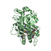
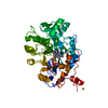
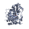
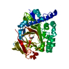
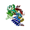
| Title | Functional and structural insights into the unusual oxyanion hole-like geometry in macrolactin acyltransferase selective for dicarboxylic acyl donors | ||||||
 Components Components | Putative beta-lactamase | ||||||
 Keywords Keywords | TRANSFERASE / acyltransferase | ||||||
| Function / homology |  Function and homology information Function and homology information: / Beta-lactamase-related / Beta-lactamase / Beta-lactamase / DD-peptidase/beta-lactamase superfamily / Beta-lactamase/transpeptidase-like / 3-Layer(aba) Sandwich / Alpha Beta Similarity search - Domain/homology | ||||||
| Biological species |  Jeotgalibacillus marinus (bacteria) Jeotgalibacillus marinus (bacteria) | ||||||
| Method |  X-RAY DIFFRACTION / SYNCHROTRON / MOLECULAR REPLACEMENT / Resolution: 2.492 Å X-RAY DIFFRACTION / SYNCHROTRON / MOLECULAR REPLACEMENT / Resolution: 2.492 Å | ||||||
 Authors Authors | Xiao, F. / Dong, S. / Feng, Y. / Li, W. | ||||||
 Citation Citation | Journal: J.Am.Chem.Soc. / Year: 2020 Title: Structural Basis of Specificity for Carboxyl-Terminated Acyl Donors in a Bacterial Acyltransferase. Authors: Xiao, F. / Dong, S. / Liu, Y. / Feng, Y. / Li, H. / Yun, C.H. / Cui, Q. / Li, W. | ||||||
| History |
|
- Structure visualization
| Structure viewer | Molecule: MolmilJmol/JSmol |
|---|
- Downloads & links
-Download
| PDBx/mmCIF format | 6kjj.cif.gz | 161.4 KB | Display | PDBx/mmCIF format |
|---|---|---|---|---|
| PDB format | pdb6kjj.ent.gz | 126 KB | Display | PDB format |
| PDBx/mmJSON format | 6kjj.json.gz | Tree view | PDBx/mmJSON format | |
| Others |  Other downloads Other downloads |
-Validation report
| Arichive directory | https://data.pdbj.org/pub/pdb/validation_reports/kj/6kjjftp://data.pdbj.org/pub/pdb/validation_reports/kj/6kjj | HTTPS FTP |
|---|
-Related structure data
| Related structure data |  6kjhSC  6kjpC  6kjqC  6kjrC  6kjtC S: Starting model for refinement C: citing same article ( |
|---|---|
| Similar structure data |

-Links
 PDBj
PDBj
- Assembly
| Deposited unit | 
| |||||||||
|---|---|---|---|---|---|---|---|---|---|---|
| 1 |
| |||||||||
| Unit cell |
| |||||||||
| Components on special symmetry positions |
|
-Components
| #1: Protein | Mass: 44223.426 Da / Num. of mol.: 1 / Mutation: S73A Source method: isolated from a genetically manipulated source Source: (gene. exp.) Jeotgalibacillus marinus (bacteria) / Gene: mlnI / Production host: |
|---|---|
| #2: Chemical | ChemComp-D9L /   Mass: 219.258 Da / Num. of mol.: 1 / Source method: obtained synthetically / Formula: C8H13NO4S / Feature type: SUBJECT OF INVESTIGATION Mass: 219.258 Da / Num. of mol.: 1 / Source method: obtained synthetically / Formula: C8H13NO4S / Feature type: SUBJECT OF INVESTIGATION |
| #3: Water | ChemComp-HOH /  Mass: 18.015 Da / Num. of mol.: 80 / Source method: isolated from a natural source / Formula: H2O Mass: 18.015 Da / Num. of mol.: 80 / Source method: isolated from a natural source / Formula: H2O |
| Has ligand of interest | Y |
-Experimental details
-Experiment
| Experiment | Method: X-RAY DIFFRACTION / Number of used crystals: 1 |
|---|
- Sample preparation
| Crystal | Density Matthews: 2.06 Å3/Da / Density % sol: 40.31 % |
|---|---|
| Crystal grow | Temperature: 291 K / Method: vapor diffusion, hanging drop Details: 0.2 M ammonium sulfate, 0.1 M Bis-Tris, pH 5.3, 25 % PEG3350 |
-Data collection
| Diffraction | Mean temperature: 77 K / Serial crystal experiment: N | ||||||||||||||||||||||||||||||
|---|---|---|---|---|---|---|---|---|---|---|---|---|---|---|---|---|---|---|---|---|---|---|---|---|---|---|---|---|---|---|---|
| Diffraction source | Source: SYNCHROTRON / Site: SSRF  / Beamline: BL17U1 / Wavelength: 0.979 Å / Beamline: BL17U1 / Wavelength: 0.979 Å | ||||||||||||||||||||||||||||||
| Detector | Type: ADSC QUANTUM 315r / Detector: CCD / Date: Jun 2, 2017 | ||||||||||||||||||||||||||||||
| Radiation | Protocol: SINGLE WAVELENGTH / Monochromatic (M) / Laue (L): M / Scattering type: x-ray | ||||||||||||||||||||||||||||||
| Radiation wavelength | Wavelength: 0.979 Å / Relative weight: 1 | ||||||||||||||||||||||||||||||
| Reflection | Resolution: 2.49→117.99 Å / Num. obs: 13437 / % possible obs: 99.9 % / Redundancy: 13.7 % / CC1/2: 0.998 / Rmerge(I) obs: 0.098 / Rpim(I) all: 0.028 / Rrim(I) all: 0.102 / Net I/σ(I): 22.5 / Num. measured all: 184221 | ||||||||||||||||||||||||||||||
| Reflection shell | Diffraction-ID: 1
|
- Processing
| Software |
| ||||||||||||||||||||||||||||||||||||||||||||||||||||||||||||||||||||||||||||||||||||||||||||||||||||||||||||||||||||||||||||||||||||||||||||||||||||||
|---|---|---|---|---|---|---|---|---|---|---|---|---|---|---|---|---|---|---|---|---|---|---|---|---|---|---|---|---|---|---|---|---|---|---|---|---|---|---|---|---|---|---|---|---|---|---|---|---|---|---|---|---|---|---|---|---|---|---|---|---|---|---|---|---|---|---|---|---|---|---|---|---|---|---|---|---|---|---|---|---|---|---|---|---|---|---|---|---|---|---|---|---|---|---|---|---|---|---|---|---|---|---|---|---|---|---|---|---|---|---|---|---|---|---|---|---|---|---|---|---|---|---|---|---|---|---|---|---|---|---|---|---|---|---|---|---|---|---|---|---|---|---|---|---|---|---|---|---|---|---|---|
| Refinement | Method to determine structure: MOLECULAR REPLACEMENT Starting model: 6KJH Resolution: 2.492→39.165 Å / SU ML: 0.24 / Cross valid method: THROUGHOUT / σ(F): 1.34 / Phase error: 23.34
| ||||||||||||||||||||||||||||||||||||||||||||||||||||||||||||||||||||||||||||||||||||||||||||||||||||||||||||||||||||||||||||||||||||||||||||||||||||||
| Solvent computation | Shrinkage radii: 0.9 Å / VDW probe radii: 1.11 Å | ||||||||||||||||||||||||||||||||||||||||||||||||||||||||||||||||||||||||||||||||||||||||||||||||||||||||||||||||||||||||||||||||||||||||||||||||||||||
| Displacement parameters | Biso max: 118.2 Å2 / Biso mean: 37.4035 Å2 / Biso min: 10.52 Å2 | ||||||||||||||||||||||||||||||||||||||||||||||||||||||||||||||||||||||||||||||||||||||||||||||||||||||||||||||||||||||||||||||||||||||||||||||||||||||
| Refinement step | Cycle: final / Resolution: 2.492→39.165 Å
| ||||||||||||||||||||||||||||||||||||||||||||||||||||||||||||||||||||||||||||||||||||||||||||||||||||||||||||||||||||||||||||||||||||||||||||||||||||||
| LS refinement shell | Refine-ID: X-RAY DIFFRACTION / Rfactor Rfree error: 0
| ||||||||||||||||||||||||||||||||||||||||||||||||||||||||||||||||||||||||||||||||||||||||||||||||||||||||||||||||||||||||||||||||||||||||||||||||||||||
| Refinement TLS params. | Method: refined / Refine-ID: X-RAY DIFFRACTION
| ||||||||||||||||||||||||||||||||||||||||||||||||||||||||||||||||||||||||||||||||||||||||||||||||||||||||||||||||||||||||||||||||||||||||||||||||||||||
| Refinement TLS group |
|
