| 登録情報 | データベース: PDB / ID: 1jwg
|
|---|
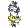
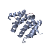
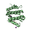
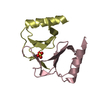
| タイトル | VHS Domain of human GGA1 complexed with cation-independent M6PR C-terminal Peptide |
|---|
 要素 要素 | - ADP-ribosylation factor binding protein GGA1
- Cation-independent mannose-6-phosphate receptor
|
|---|
 キーワード キーワード | PROTEIN TRANSPORT/PROTEIN BINDING / SUPER HELIX / PROTEIN-PEPTIDE COMPLEX / PROTEIN TRANSPORT-PROTEIN BINDING COMPLEX |
|---|
| 機能・相同性 |  機能・相同性情報 機能・相同性情報
protein localization to ciliary membrane / clathrin coat / Retrograde transport at the Trans-Golgi-Network / response to tetrachloromethane / insulin-like growth factor receptor activity / retromer complex binding / insulin-like growth factor binding / insulin-like growth factor II binding / trans-Golgi network transport vesicle / Golgi to plasma membrane transport ...protein localization to ciliary membrane / clathrin coat / Retrograde transport at the Trans-Golgi-Network / response to tetrachloromethane / insulin-like growth factor receptor activity / retromer complex binding / insulin-like growth factor binding / insulin-like growth factor II binding / trans-Golgi network transport vesicle / Golgi to plasma membrane transport / host-mediated activation of viral process / retinoic acid binding / Golgi to plasma membrane protein transport / retrograde transport, endosome to Golgi / TBC/RABGAPs / protein localization to cell surface / lysosomal transport / Golgi Associated Vesicle Biogenesis / nuclear envelope lumen / D-mannose binding / endocytic vesicle / G-protein alpha-subunit binding / animal organ regeneration / response to retinoic acid / transport vesicle / phosphatidylinositol binding / receptor-mediated endocytosis / secretory granule membrane / ubiquitin binding / trans-Golgi network membrane / post-embryonic development / intracellular protein transport / phosphoprotein binding / trans-Golgi network / clathrin-coated endocytic vesicle membrane / protein catabolic process / liver development / small GTPase binding / positive regulation of protein catabolic process / Cargo recognition for clathrin-mediated endocytosis / Clathrin-mediated endocytosis / intracellular protein localization / late endosome / signaling receptor activity / early endosome membrane / spermatogenesis / early endosome / endosome membrane / endosome / positive regulation of apoptotic process / G protein-coupled receptor signaling pathway / Amyloid fiber formation / Golgi membrane / focal adhesion / intracellular membrane-bounded organelle / Neutrophil degranulation / perinuclear region of cytoplasm / enzyme binding / cell surface / Golgi apparatus / signal transduction / protein-containing complex / extracellular exosome / nucleoplasm / identical protein binding / membrane / plasma membrane / cytosol類似検索 - 分子機能 ADP-ribosylation factor-binding protein GGA3 / N-terminal extension of GAT domain / N-terminal extension of GAT domain / GAT domain / GAT domain superfamily / GAT domain / GAT domain profile. / Cation-independent mannose-6-phosphate receptor repeat / Cation-independent mannose-6-phosphate receptor repeat / Cation-independent mannose-6-phosphate receptor repeat ...ADP-ribosylation factor-binding protein GGA3 / N-terminal extension of GAT domain / N-terminal extension of GAT domain / GAT domain / GAT domain superfamily / GAT domain / GAT domain profile. / Cation-independent mannose-6-phosphate receptor repeat / Cation-independent mannose-6-phosphate receptor repeat / Cation-independent mannose-6-phosphate receptor repeat / VHS domain / VHS domain / VHS domain profile. / Domain present in VPS-27, Hrs and STAM / Gamma-adaptin ear (GAE) domain / Gamma-adaptin ear (GAE) domain profile. / Serine Threonine Protein Phosphatase 5, Tetratricopeptide repeat - #90 / Mannose-6-phosphate receptor binding domain superfamily / MRH domain / MRH domain profile. / Clathrin adaptor, alpha/beta/gamma-adaptin, appendage, Ig-like subdomain / Adaptin C-terminal domain / Adaptin C-terminal domain / Clathrin adaptor, appendage, Ig-like subdomain superfamily / ENTH/VHS / Fibronectin type II domain / Fibronectin type II domain superfamily / Fibronectin type II domain / Fibronectin type-II collagen-binding domain signature. / Fibronectin type-II collagen-binding domain profile. / Fibronectin type 2 domain / Kringle-like fold / Serine Threonine Protein Phosphatase 5, Tetratricopeptide repeat / Alpha Horseshoe / Mainly Alpha類似検索 - ドメイン・相同性 IODIDE ION / Cation-independent mannose-6-phosphate receptor / ADP-ribosylation factor-binding protein GGA1類似検索 - 構成要素 |
|---|
| 生物種 |  Homo sapiens (ヒト) Homo sapiens (ヒト) |
|---|
| 手法 |  X線回折 / シンクロトロン / 分子置換 / 解像度: 2 Å X線回折 / シンクロトロン / 分子置換 / 解像度: 2 Å |
|---|
 データ登録者 データ登録者 | Shiba, T. / Takatsu, H. / Nogi, T. / Matsugaki, N. / Kawasaki, M. / Igarashi, N. / Suzuki, M. / Kato, R. / Earnest, T. / Nakayama, K. / Wakatsuki, S. |
|---|
 引用 引用 | ジャーナル: Nature / 年: 2002
タイトル: Structural basis for recognition of acidic-cluster dileucine sequence by GGA1.
著者: Shiba, T. / Takatsu, H. / Nogi, T. / Matsugaki, N. / Kawasaki, M. / Igarashi, N. / Suzuki, M. / Kato, R. / Earnest, T. / Nakayama, K. / Wakatsuki, S. |
|---|
| 履歴 | | 登録 | 2001年9月4日 | 登録サイト: RCSB / 処理サイト: PDBJ |
|---|
| 改定 1.0 | 2002年3月6日 | Provider: repository / タイプ: Initial release |
|---|
| 改定 1.1 | 2008年4月27日 | Group: Version format compliance |
|---|
| 改定 1.2 | 2011年7月13日 | Group: Version format compliance |
|---|
| 改定 1.3 | 2023年10月25日 | Group: Data collection / Database references ...Data collection / Database references / Derived calculations / Refinement description
カテゴリ: chem_comp_atom / chem_comp_bond ...chem_comp_atom / chem_comp_bond / database_2 / pdbx_initial_refinement_model / struct_site
Item: _database_2.pdbx_DOI / _database_2.pdbx_database_accession ..._database_2.pdbx_DOI / _database_2.pdbx_database_accession / _struct_site.pdbx_auth_asym_id / _struct_site.pdbx_auth_comp_id / _struct_site.pdbx_auth_seq_id |
|---|
| 改定 1.4 | 2024年10月9日 | Group: Structure summary
カテゴリ: pdbx_entry_details / pdbx_modification_feature |
|---|
|
|---|
 ムービー
ムービー コントローラー


 データを開く
 基本情報
基本情報 構造の表示
構造の表示 ダウンロードとリンク
 その他のダウンロード
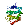


 PDBj
PDBj


 集合体
集合体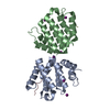


 分子量: 126.904 Da / 分子数: 6 / 由来タイプ: 合成 / 式: I
 分子量: 18.015 Da / 分子数: 198 / 由来タイプ: 天然 / 式: H2O
 試料調製
試料調製 / ビームライン: 5.0.2 / 波長: 1 Å
 解析
解析