 Movie
Movie Controller

[English] 日本語
 Yorodumi
- PDB-1jwd: Ca2+-induced Structural Changes in Calcyclin: High-resolution Sol... -
+ Open data

- Basic information
| Entry | Database: PDB / ID: 1jwd | ||||||
|---|---|---|---|---|---|---|---|
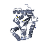
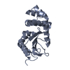
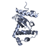
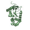
| Title | Ca2+-induced Structural Changes in Calcyclin: High-resolution Solution Structure of Ca2+-bound Calcyclin. | ||||||
 Components Components | Calcyclin | ||||||
 Keywords Keywords | METAL BINDING PROTEIN / Ca(2+)-binding protein / S100 protein / EF-hand / S100A6 | ||||||
| Function / homology |  Function and homology information Function and homology informationS100 protein binding / cytoplasmic side of plasma membrane / calcium-dependent protein binding / nuclear envelope / calcium ion binding / perinuclear region of cytoplasm / : / cytosol Similarity search - Function | ||||||
| Biological species |  | ||||||
| Method | SOLUTION NMR / distance geometry, restrained molecular dynamics | ||||||
 Authors Authors | Maler, L. / Sastry, M. / Chazin, W.J. | ||||||
 Citation Citation | Journal: J.Mol.Biol. / Year: 2002 Title: A structural basis for S100 protein specificity derived from comparative analysis of apo and Ca(2+)-calcyclin Authors: Maler, L. / Sastry, M. / Chazin, W.J. #1: Journal: J.Biomol.NMR / Year: 1999Title: High Resolution Solution Structure of Apo Calcyclin and Structural Variations in the S100 Family of Calcium-binding Proteins. Authors: Maler, L. / Potts, B.C.M. / Chazin, W.J. #2: Journal: Structure / Year: 1998Title: The Three-dimenisonal Structure of Ca(2+)-bound Calcyclin: Implications for Ca(2+)-signal Transduction by S100 Proteins. Authors: Sastry, M. / Ketchem, R.R. / Crescenzi, O. / Weber, C. / Lubienski, M.J. / Hidaka, H. / Chazin, W.J. #3: Journal: Nat.Struct.Biol. / Year: 1995Title: The Structure of Calcyclin Reveals a Novel Homodimeric Fold for S100 Ca(2+)-binding Proteins. Authors: Potts, B.C. / Smith, J. / Akke, M. / Macke, T.J. / Okazaki, K. / Hidaka, H. / Chase, D.A. / Chazin, W.J. | ||||||
| History |
|
- Structure visualization
| Structure viewer | Molecule: MolmilJmol/JSmol |
|---|
- Downloads & links
-Download
| PDBx/mmCIF format | 1jwd.cif.gz | 1.2 MB | Display | PDBx/mmCIF format |
|---|---|---|---|---|
| PDB format | pdb1jwd.ent.gz | 1 MB | Display | PDB format |
| PDBx/mmJSON format | 1jwd.json.gz | Tree view | PDBx/mmJSON format | |
| Others |  Other downloads Other downloads |
-Validation report
| Arichive directory | https://data.pdbj.org/pub/pdb/validation_reports/jw/1jwdftp://data.pdbj.org/pub/pdb/validation_reports/jw/1jwd | HTTPS FTP |
|---|
-Related structure data
| Related structure data | |
|---|---|
| Similar structure data |


-Links
 PDBj
PDBj- Assembly
| Deposited unit | 
| |||||||||
|---|---|---|---|---|---|---|---|---|---|---|
| 1 |
| |||||||||
| NMR ensembles |
|
-Components
| #1: Protein | Mass: 10167.729 Da / Num. of mol.: 2 Source method: isolated from a genetically manipulated source Source: (gene. exp.)  |
|---|
-Experimental details
-Experiment
| Experiment | Method: SOLUTION NMR | ||||||||||||||||||||||||||||||||
|---|---|---|---|---|---|---|---|---|---|---|---|---|---|---|---|---|---|---|---|---|---|---|---|---|---|---|---|---|---|---|---|---|---|
| NMR experiment |
| ||||||||||||||||||||||||||||||||
| NMR details | Text: Dimer constraints were obtained from the 3D_13C-filter,13C-edited experiment in combination with 3D_13C-separated_NOESY |
 HSQC
HSQC- Sample preparation
| Details |
| ||||||||||||||||||
|---|---|---|---|---|---|---|---|---|---|---|---|---|---|---|---|---|---|---|---|
| Sample conditions | Ionic strength: 30 mM CaCl2 / pH: 7 / Pressure: ambient / Temperature: 300 K | ||||||||||||||||||
| Crystal grow | *PLUS Method: other / Details: NMR |
-NMR measurement
| Radiation | Protocol: SINGLE WAVELENGTH / Monochromatic (M) / Laue (L): M | ||||||||||||||||||||
|---|---|---|---|---|---|---|---|---|---|---|---|---|---|---|---|---|---|---|---|---|---|
| Radiation wavelength | Relative weight: 1 | ||||||||||||||||||||
| NMR spectrometer |
|
- Processing
| NMR software |
| ||||||||||||||||||||
|---|---|---|---|---|---|---|---|---|---|---|---|---|---|---|---|---|---|---|---|---|---|
| Refinement | Method: distance geometry, restrained molecular dynamics / Software ordinal: 1 Details: The calculations were carried out using a total of 3104 distance and 294 torsion angle constraints. Starting structures were generated as monomers (one chain) with no intersubunit ...Details: The calculations were carried out using a total of 3104 distance and 294 torsion angle constraints. Starting structures were generated as monomers (one chain) with no intersubunit constraints using distance geometry followed by restrained molecular dynamics (rMD). The dimer structures were generated by rMD docking driven by the intersubunit NOEs using two arbitrarily selected starting subunit structures. Each dimer was further refined by rMD with all constraints. | ||||||||||||||||||||
| NMR representative | Selection criteria: closest to the average | ||||||||||||||||||||
| NMR ensemble | Conformer selection criteria: The program Findfam was used to establish that the number of structures required to accurately represent the ensemble was less than 22 (the number selected to represent ...Conformer selection criteria: The program Findfam was used to establish that the number of structures required to accurately represent the ensemble was less than 22 (the number selected to represent previous S100A6 ensembles). Structures were ordered by lowest restraint violations, then accepted if total molecular energy and each contributing term was within two standard deviations of the mean. The 22 structures with least restraint violations (energy penalty and magnitude of largest violation) all met these criteria. Conformers calculated total number: 100 / Conformers submitted total number: 22 |
