 Movie
Movie Controller

[English] 日本語
 Yorodumi
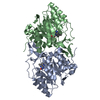
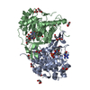
- PDB-1jrb: The P56A mutant of Lactococcus lactis dihydroorotate dehydrogenase A -
+ Open data

- Basic information
| Entry | Database: PDB / ID: 1jrb | ||||||
|---|---|---|---|---|---|---|---|
| Title | The P56A mutant of Lactococcus lactis dihydroorotate dehydrogenase A | ||||||
 Components Components | dihydroorotate dehydrogenase A | ||||||
 Keywords Keywords | OXIDOREDUCTASE / Homodimer / alpha-beta barrel / flavoprotein / orotate complex / mutant enzyme | ||||||
| Function / homology |  Function and homology information Function and homology informationdihydroorotate dehydrogenase (fumarate) / dihydroorotate dehydrogenase (fumarate) activity / 'de novo' UMP biosynthetic process / 'de novo' pyrimidine nucleobase biosynthetic process / cytoplasm Similarity search - Function | ||||||
| Biological species |  Lactococcus lactis (lactic acid bacteria) Lactococcus lactis (lactic acid bacteria) | ||||||
| Method |  X-RAY DIFFRACTION / SYNCHROTRON / MOLECULAR REPLACEMENT / Resolution: 1.9 Å X-RAY DIFFRACTION / SYNCHROTRON / MOLECULAR REPLACEMENT / Resolution: 1.9 Å | ||||||
 Authors Authors | Norager, S. / Arent, S. / Bjornberg, O. / Ottosen, M. / Lo Leggio, L. / Jensen, K.F. / Larsen, S. | ||||||
 Citation Citation | Journal: J.Biol.Chem. / Year: 2003 Title: Lactococcus lactis dihydroorotate dehydrogenase A mutants reveal important facets of the enzymatic function Authors: Norager, S. / Arent, S. / Bjornberg, O. / Ottosen, M. / Lo Leggio, L. / Jensen, K.F. / Larsen, S. #1: Journal: Biochemistry / Year: 1997Title: Active site of dihydroorotate dehydrogenase A from Lactococcus lactis investigated by chemical modification and mutagenesis Authors: Bjornberg, O. / Rowland, P. / Larsen, S. / Jensen, K.F. #2: Journal: Protein Sci. / Year: 1998Title: The crystal structure of Lactococcus lactis dihydroorotate dehydrogenase A complexed with the enzyme reaction product throws light on its enzymatic function Authors: Rowland, P. / Bjornberg, O. / Nielsen, F.S. / Jensen, K.F. / Larsen, S. #3: Journal: Structure / Year: 1997Title: The crystal structure of the flavin containing enzyme dihydroorotate dehydrogenase A from Lactococcus lactis Authors: Rowland, P. / Nielsen, F.S. / Jensen, K.F. / Larsen, S. #4: Journal: Protein Sci. / Year: 1996Title: Purification and characterisation of dihydroorotate dehydrogenase A from Lactococcus lactis, crystallisation and preliminary X-ray diffraction studies of the enzyme Authors: Nielsen, F.S. / Rowland, P. / Larsen, S. / Jensen, K.F. | ||||||
| History |
|
- Structure visualization
| Structure viewer | Molecule: MolmilJmol/JSmol |
|---|
- Downloads & links
-Download
| PDBx/mmCIF format | 1jrb.cif.gz | 142.9 KB | Display | PDBx/mmCIF format |
|---|---|---|---|---|
| PDB format | pdb1jrb.ent.gz | 110.2 KB | Display | PDB format |
| PDBx/mmJSON format | 1jrb.json.gz | Tree view | PDBx/mmJSON format | |
| Others |  Other downloads Other downloads |
-Validation report
| Summary document | 1jrb_validation.pdf.gz | 1 MB | Display | wwPDB validaton report |
|---|---|---|---|---|
| Full document | 1jrb_full_validation.pdf.gz | 1.1 MB | Display | |
| Data in XML | 1jrb_validation.xml.gz | 31.4 KB | Display | |
| Data in CIF | 1jrb_validation.cif.gz | 43.5 KB | Display | |
| Arichive directory | https://data.pdbj.org/pub/pdb/validation_reports/jr/1jrbftp://data.pdbj.org/pub/pdb/validation_reports/jr/1jrb | HTTPS FTP |
-Related structure data
| Related structure data | 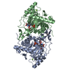 1jqvC 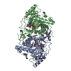 1jqxC  1jrcC  1jubC  1jueC  1ovdC 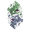 2dorS S: Starting model for refinement C: citing same article ( |
|---|---|
| Similar structure data |

-Links
 PDBj
PDBj- Assembly
| Deposited unit | 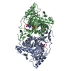
| ||||||||
|---|---|---|---|---|---|---|---|---|---|
| 1 |
| ||||||||
| Unit cell |
| ||||||||
| Details | The assymetric unit contains the biological homodimer. |
-Components
| #1: Protein | Mass: 34216.133 Da / Num. of mol.: 2 / Mutation: P56A Source method: isolated from a genetically manipulated source Source: (gene. exp.) Lactococcus lactis (lactic acid bacteria)Plasmid: pUHE23 / Production host: References: UniProt: P54321, UniProt: A2RJT9*PLUS, EC: 1.3.3.1 #2: Chemical |   Mass: 456.344 Da / Num. of mol.: 2 / Source method: obtained synthetically / Formula: C17H21N4O9P Mass: 456.344 Da / Num. of mol.: 2 / Source method: obtained synthetically / Formula: C17H21N4O9P#3: Chemical | 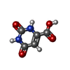  Mass: 156.096 Da / Num. of mol.: 2 / Source method: obtained synthetically / Formula: C5H4N2O4 Mass: 156.096 Da / Num. of mol.: 2 / Source method: obtained synthetically / Formula: C5H4N2O4#4: Water | ChemComp-HOH / |  Mass: 18.015 Da / Num. of mol.: 358 / Source method: isolated from a natural source / Formula: H2O Mass: 18.015 Da / Num. of mol.: 358 / Source method: isolated from a natural source / Formula: H2O |
|---|
-Experimental details
-Experiment
| Experiment | Method: X-RAY DIFFRACTION / Number of used crystals: 1 |
|---|
- Sample preparation
| Crystal | Density Matthews: 2.72 Å3/Da / Density % sol: 54.79 % |
|---|---|
| Crystal grow | Temperature: 293 K / Method: vapor diffusion, hanging drop Details: PEG 6K, Na-acetate, TRIS-HCl, VAPOR DIFFUSION, HANGING DROP, temperature 293K |
-Data collection
| Diffraction | Mean temperature: 120 K |
|---|---|
| Diffraction source | Source: SYNCHROTRON / Site: MAX II  / Beamline: I711 / Wavelength: 0.992 Å / Beamline: I711 / Wavelength: 0.992 Å |
| Detector | Type: MARRESEARCH / Detector: IMAGE PLATE / Date: Mar 17, 1999 |
| Radiation | Monochromator: Si(111) single crystal / Protocol: SINGLE WAVELENGTH / Monochromatic (M) / Laue (L): M / Scattering type: x-ray |
| Radiation wavelength | Wavelength: 0.992 Å / Relative weight: 1 |
| Reflection | Resolution: 1.93→20 Å / Num. all: 452819 / Num. obs: 57637 / % possible obs: 93.6 % / Observed criterion σ(F): 0 / Observed criterion σ(I): 0 / Redundancy: 3.8 % / Biso Wilson estimate: 14.4 Å2 / Rmerge(I) obs: 0.046 / Net I/σ(I): 29.9 |
| Reflection shell | Resolution: 1.93→1.97 Å / Rmerge(I) obs: 0.187 / Mean I/σ(I) obs: 6.2 / Num. unique all: 2390 / % possible all: 82.2 |
- Processing
| Software |
| ||||||||||||||||||||||||||||||||||||||||||||||||
|---|---|---|---|---|---|---|---|---|---|---|---|---|---|---|---|---|---|---|---|---|---|---|---|---|---|---|---|---|---|---|---|---|---|---|---|---|---|---|---|---|---|---|---|---|---|---|---|---|---|
| Refinement | Method to determine structure: MOLECULAR REPLACEMENT Starting model: Lactococcus lactis DHODA, PDB ID 2DOR Resolution: 1.9→20 Å / Isotropic thermal model: isotropic / Cross valid method: THROUGHOUT / σ(F): 0 / σ(I): 0 / Stereochemistry target values: Engh&Huber Details: Data obtained after the termination of this structure has enabled us to identifie to MG-ions in the lactococcus lactis DHODA. These are also present in the P56A mutant but have been refined ...Details: Data obtained after the termination of this structure has enabled us to identifie to MG-ions in the lactococcus lactis DHODA. These are also present in the P56A mutant but have been refined as water molecules 11 and 23. The close contacts with water molecules are due to disorderd residues refined with low occupation.
| ||||||||||||||||||||||||||||||||||||||||||||||||
| Displacement parameters | Biso mean: 22.97 Å2 | ||||||||||||||||||||||||||||||||||||||||||||||||
| Refinement step | Cycle: LAST / Resolution: 1.9→20 Å
| ||||||||||||||||||||||||||||||||||||||||||||||||
| Refine LS restraints |
| ||||||||||||||||||||||||||||||||||||||||||||||||
| Refine LS restraints NCS | NCS model details: RESTRAIND | ||||||||||||||||||||||||||||||||||||||||||||||||
| LS refinement shell | Resolution: 1.934→2.025 Å
|
