 Movie
Movie Controller

[English] 日本語
 Yorodumi
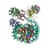
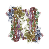
- PDB-1jqn: Crystal structure of E.coli phosphoenolpyruvate carboxylase in co... -
+ Open data

- Basic information
| Entry | Database: PDB / ID: 1jqn | ||||||
|---|---|---|---|---|---|---|---|
| Title | Crystal structure of E.coli phosphoenolpyruvate carboxylase in complex with Mn2+ and DCDP | ||||||
 Components Components | phosphoenolpyruvate carboxylase | ||||||
 Keywords Keywords | LYASE / BETA BARREL / Mn2+ and DCDP COMPLEX | ||||||
| Function / homology |  Function and homology information Function and homology informationphosphoenolpyruvate carboxylase / phosphoenolpyruvate carboxylase activity / oxaloacetate metabolic process / carbon fixation / tricarboxylic acid cycle / gluconeogenesis / protein homotetramerization / magnesium ion binding / identical protein binding / cytosol Similarity search - Function | ||||||
| Biological species |  | ||||||
| Method |  X-RAY DIFFRACTION / SYNCHROTRON / MOLECULAR REPLACEMENT / Resolution: 2.35 Å X-RAY DIFFRACTION / SYNCHROTRON / MOLECULAR REPLACEMENT / Resolution: 2.35 Å | ||||||
 Authors Authors | Matsumura, H. / Kai, Y. | ||||||
 Citation Citation | Journal: Structure / Year: 2002 Title: Crystal structures of C4 form maize and quaternary complex of E. coli phosphoenolpyruvate carboxylases. Authors: Matsumura, H. / Xie, Y. / Shirakata, S. / Inoue, T. / Yoshinaga, T. / Ueno, Y. / Izui, K. / Kai, Y. #1: Journal: FEBS Lett. / Year: 1999Title: Plausible phosphoenolpyruvate binding site revealed by 2.6 A structure of Mn2+-bound phosphoenolpyruvate carboxylase from Escherichia coli. Authors: Matsumura, H. / Terada, M. / Shirakata, S. / Inoue, T. / Yoshinaga, T. / Izui, K. / Kai, Y. #2: Journal: Proc.Natl.Acad.Sci.USA / Year: 1999Title: Three-dimensional structure of phosphoenolpyruvate carboxylase: a proposed mechanism for allosteric inhibition Authors: Kai, Y. / Matsumura, H. / Inoue, T. / Terada, K. / Nagara, Y. / Yoshinaga, T. / Kihara, A. / Tsumura, K. / Izui, K. #3: Journal: Acta Crystallogr.,Sect.D / Year: 1999Title: Crystallization and preliminary x-ray diffraction studies of C4-form phosphoenolpyruvate carboxylase from maize diffraction studies of C4-form phosphoenolpyruvate carboxylase from maize. Authors: Matsumura, H. / Nagata, T. / Terada, M. / Shirakata, S. / Inoue, T. / Yoshinaga, T. / Ueno, Y. / Saze, H. / Izui, K. / Kai, Y. | ||||||
| History |
|
- Structure visualization
| Structure viewer | Molecule: MolmilJmol/JSmol |
|---|
- Downloads & links
-Download
| PDBx/mmCIF format | 1jqn.cif.gz | 185 KB | Display | PDBx/mmCIF format |
|---|---|---|---|---|
| PDB format | pdb1jqn.ent.gz | 147 KB | Display | PDB format |
| PDBx/mmJSON format | 1jqn.json.gz | Tree view | PDBx/mmJSON format | |
| Others |  Other downloads Other downloads |
-Validation report
| Arichive directory | https://data.pdbj.org/pub/pdb/validation_reports/jq/1jqnftp://data.pdbj.org/pub/pdb/validation_reports/jq/1jqn | HTTPS FTP |
|---|
-Related structure data


-Links
 PDBj
PDBj

- Assembly
| Deposited unit | 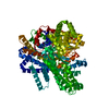
| ||||||||
|---|---|---|---|---|---|---|---|---|---|
| 1 | 
| ||||||||
| Unit cell |
| ||||||||
| Details | The biological assembly is a tetramer generated from monomer in the assymmetric unit by D2 symmetry. |
-Components
| #1: Protein | Mass: 99175.492 Da / Num. of mol.: 1 Source method: isolated from a genetically manipulated source Source: (gene. exp.) References: UniProt: P00864, phosphoenolpyruvate carboxylase |
|---|---|
| #2: Chemical | ChemComp-MN /   Mass: 54.938 Da / Num. of mol.: 1 / Source method: obtained synthetically / Formula: Mn Mass: 54.938 Da / Num. of mol.: 1 / Source method: obtained synthetically / Formula: Mn |
| #3: Chemical | ChemComp-ASP /   Type: L-peptide linking / Mass: 133.103 Da / Num. of mol.: 1 / Source method: obtained synthetically / Formula: C4H7NO4 Type: L-peptide linking / Mass: 133.103 Da / Num. of mol.: 1 / Source method: obtained synthetically / Formula: C4H7NO4 |
| #4: Chemical | ChemComp-DCO / 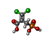  Mass: 234.959 Da / Num. of mol.: 1 / Source method: obtained synthetically / Formula: C4H5Cl2O5P Mass: 234.959 Da / Num. of mol.: 1 / Source method: obtained synthetically / Formula: C4H5Cl2O5P |
| #5: Water | ChemComp-HOH /  Mass: 18.015 Da / Num. of mol.: 194 / Source method: isolated from a natural source / Formula: H2O Mass: 18.015 Da / Num. of mol.: 194 / Source method: isolated from a natural source / Formula: H2O |
-Experimental details
-Experiment
| Experiment | Method: X-RAY DIFFRACTION / Number of used crystals: 1 |
|---|
- Sample preparation
| Crystal | Density Matthews: 3.08 Å3/Da / Density % sol: 60.03 % | ||||||||||||||||||||||||||||||||||||||||||||||||||||||||||||||||||||||||||||||||||||
|---|---|---|---|---|---|---|---|---|---|---|---|---|---|---|---|---|---|---|---|---|---|---|---|---|---|---|---|---|---|---|---|---|---|---|---|---|---|---|---|---|---|---|---|---|---|---|---|---|---|---|---|---|---|---|---|---|---|---|---|---|---|---|---|---|---|---|---|---|---|---|---|---|---|---|---|---|---|---|---|---|---|---|---|---|---|
| Crystal grow | Temperature: 293 K / Method: vapor diffusion, hanging drop / pH: 8 Details: PEG300, Tris-HCl, MnCl2, DCDP, pH 8.0, VAPOR DIFFUSION, HANGING DROP, temperature 293K | ||||||||||||||||||||||||||||||||||||||||||||||||||||||||||||||||||||||||||||||||||||
| Crystal grow | *PLUS pH: 7.4 | ||||||||||||||||||||||||||||||||||||||||||||||||||||||||||||||||||||||||||||||||||||
| Components of the solutions | *PLUS
|
-Data collection
| Diffraction | Mean temperature: 293 K |
|---|---|
| Diffraction source | Source: SYNCHROTRON / Site: Photon Factory  / Beamline: BL-18B / Wavelength: 1 Å / Beamline: BL-18B / Wavelength: 1 Å |
| Detector | Type: WEISSENBERG / Detector: DIFFRACTOMETER / Date: Nov 24, 1999 |
| Radiation | Monochromator: Si 111 CHANNEL / Protocol: SINGLE WAVELENGTH / Monochromatic (M) / Laue (L): M / Scattering type: x-ray |
| Radiation wavelength | Wavelength: 1 Å / Relative weight: 1 |
| Reflection | Resolution: 2.35→39.5 Å / Num. all: 233027 / Num. obs: 45789 / % possible obs: 89.2 % / Observed criterion σ(I): 11.3 / Rmerge(I) obs: 0.058 |
| Reflection shell | Resolution: 2.35→2.43 Å / Rmerge(I) obs: 0.302 / % possible all: 61.9 |
| Reflection | *PLUS Num. measured all: 233027 |
- Processing
| Software |
| |||||||||||||||||||||||||
|---|---|---|---|---|---|---|---|---|---|---|---|---|---|---|---|---|---|---|---|---|---|---|---|---|---|---|
| Refinement | Method to determine structure: MOLECULAR REPLACEMENT / Resolution: 2.35→39.5 Å / σ(F): 0 / Stereochemistry target values: Engh & Huber
| |||||||||||||||||||||||||
| Refine analyze | Luzzati coordinate error obs: 0.31 Å / Luzzati d res low obs: 5 Å / Luzzati sigma a obs: 0.56 Å | |||||||||||||||||||||||||
| Refinement step | Cycle: LAST / Resolution: 2.35→39.5 Å
| |||||||||||||||||||||||||
| Refine LS restraints |
| |||||||||||||||||||||||||
| LS refinement shell | Resolution: 2.35→2.5 Å / Rfactor Rfree error: 0.021
| |||||||||||||||||||||||||
| Refinement | *PLUS % reflection Rfree: 5 % | |||||||||||||||||||||||||
| Solvent computation | *PLUS | |||||||||||||||||||||||||
| Displacement parameters | *PLUS | |||||||||||||||||||||||||
| Refine LS restraints | *PLUS
| |||||||||||||||||||||||||
| LS refinement shell | *PLUS Lowest resolution: 2.5 Å |
