 Movie
Movie Controller

[English] 日本語
 Yorodumi
- PDB-1jqo: Crystal structure of C4-form phosphoenolpyruvate carboxylase from... -
+ Open data

- Basic information
| Entry | Database: PDB / ID: 1jqo | ||||||
|---|---|---|---|---|---|---|---|
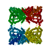
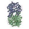
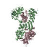
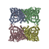
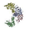
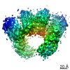
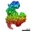
| Title | Crystal structure of C4-form phosphoenolpyruvate carboxylase from maize | ||||||
 Components Components | phosphoenolpyruvate carboxylase | ||||||
 Keywords Keywords | LYASE / BETA BARREL / Carbon dioxide fixation | ||||||
| Function / homology |  Function and homology information Function and homology informationresponse to cytokinin / phosphoenolpyruvate carboxylase / phosphoenolpyruvate carboxylase activity / response to nitrate / leaf development / response to ammonium ion / carbon fixation / tricarboxylic acid cycle / photosynthesis / cytosol Similarity search - Function | ||||||
| Biological species |  | ||||||
| Method |  X-RAY DIFFRACTION / SYNCHROTRON / MOLECULAR REPLACEMENT / Resolution: 3 Å X-RAY DIFFRACTION / SYNCHROTRON / MOLECULAR REPLACEMENT / Resolution: 3 Å | ||||||
 Authors Authors | Matsumura, H. / Kai, Y. | ||||||
 Citation Citation | Journal: Structure / Year: 2002 Title: Crystal structures of C4 form maize and quaternary complex of E. coli phosphoenolpyruvate carboxylases. Authors: Matsumura, H. / Xie, Y. / Shirakata, S. / Inoue, T. / Yoshinaga, T. / Ueno, Y. / Izui, K. / Kai, Y. #1: Journal: FEBS Lett. / Year: 1999Title: Plausible phosphoenolpyruvate binding site revealed by 2.6 A structure of Mn2+-bound phosphoenolpyruvate carboxylase from Escherichia coli. Authors: Matsumura, H. / Terada, M. / Shirakata, S. / Inoue, T. / Yoshinaga, T. / Izui, K. / Kai, Y. #2: Journal: Proc.Natl.Acad.Sci.USA / Year: 1999Title: Three-dimensional structure of phosphoenolpyruvate carboxylase: a proposed mechanism for allosteric inhibition Authors: Kai, Y. / Matsumura, H. / Inoue, T. / Terada, K. / Nagara, Y. / Yoshinaga, T. / Kihara, A. / Tsumura, K. / Izui, K. #3: Journal: Acta Crystallogr.,Sect.D / Year: 1999Title: Crystallization and preliminary x-ray diffraction studies of C4-form phosphoenolpyruvate carboxylase from maize diffraction studies of C4-form phosphoenolpyruvate carboxylase from maize. Authors: Matsumura, H. / Nagata, T. / Terada, M. / Shirakata, S. / Inoue, T. / Yoshinaga, T. / Ueno, Y. / Saze, H. / Izui, K. / Kai, Y. | ||||||
| History |
|
- Structure visualization
| Structure viewer | Molecule: MolmilJmol/JSmol |
|---|
- Downloads & links
-Download
| PDBx/mmCIF format | 1jqo.cif.gz | 358.1 KB | Display | PDBx/mmCIF format |
|---|---|---|---|---|
| PDB format | pdb1jqo.ent.gz | 291 KB | Display | PDB format |
| PDBx/mmJSON format | 1jqo.json.gz | Tree view | PDBx/mmJSON format | |
| Others |  Other downloads Other downloads |
-Validation report
| Arichive directory | https://data.pdbj.org/pub/pdb/validation_reports/jq/1jqoftp://data.pdbj.org/pub/pdb/validation_reports/jq/1jqo | HTTPS FTP |
|---|
-Related structure data

-Links
 PDBj
PDBj
- Assembly
| Deposited unit | 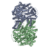
| ||||||||
|---|---|---|---|---|---|---|---|---|---|
| 1 | 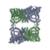
| ||||||||
| 2 |
| ||||||||
| Unit cell |
| ||||||||
| Details | The biological assembly is a tetramer generated from dimer in the assymmetric unit by two fole axis. |
-Components
| #1: Protein | Mass: 109436.008 Da / Num. of mol.: 2 / Source method: isolated from a natural source / Source: (natural) References: UniProt: P04711, phosphoenolpyruvate carboxylase #2: Chemical |   Mass: 96.063 Da / Num. of mol.: 2 / Source method: obtained synthetically / Formula: SO4 Mass: 96.063 Da / Num. of mol.: 2 / Source method: obtained synthetically / Formula: SO4 |
|---|
-Experimental details
-Experiment
| Experiment | Method: X-RAY DIFFRACTION / Number of used crystals: 2 |
|---|
- Sample preparation
| Crystal | Density Matthews: 4.02 Å3/Da / Density % sol: 69.38 % | |||||||||||||||||||||||||||||||||||||||||||||||||||||||||||||||
|---|---|---|---|---|---|---|---|---|---|---|---|---|---|---|---|---|---|---|---|---|---|---|---|---|---|---|---|---|---|---|---|---|---|---|---|---|---|---|---|---|---|---|---|---|---|---|---|---|---|---|---|---|---|---|---|---|---|---|---|---|---|---|---|---|
| Crystal grow | Temperature: 293 K / Method: vapor diffusion, hanging drop / pH: 7.5 Details: PEG8000, Tris-HCl, LiSO4, pH 7.5, VAPOR DIFFUSION, HANGING DROP, temperature 293K | |||||||||||||||||||||||||||||||||||||||||||||||||||||||||||||||
| Crystal grow | *PLUS Method: vapor diffusion, sitting drop | |||||||||||||||||||||||||||||||||||||||||||||||||||||||||||||||
| Components of the solutions | *PLUS
|
-Data collection
| Diffraction | Mean temperature: 293 K |
|---|---|
| Diffraction source | Source: SYNCHROTRON / Site: SPring-8  / Beamline: BL41XU / Wavelength: 0.708 Å / Beamline: BL41XU / Wavelength: 0.708 Å |
| Detector | Type: FUJI / Detector: IMAGE PLATE / Date: Mar 25, 1999 |
| Radiation | Monochromator: Si 111 CHANNEL / Protocol: SINGLE WAVELENGTH / Monochromatic (M) / Laue (L): M / Scattering type: x-ray |
| Radiation wavelength | Wavelength: 0.708 Å / Relative weight: 1 |
| Reflection | Resolution: 3→86.2 Å / Num. all: 266008 / Num. obs: 58078 / % possible obs: 80.4 % / Observed criterion σ(I): 7.4 / Rmerge(I) obs: 0.091 |
| Reflection shell | Resolution: 3→3.11 Å / Rmerge(I) obs: 0.309 / % possible all: 57.7 |
| Reflection | *PLUS Num. measured all: 266008 |
- Processing
| Software |
| |||||||||||||||||||||||||
|---|---|---|---|---|---|---|---|---|---|---|---|---|---|---|---|---|---|---|---|---|---|---|---|---|---|---|
| Refinement | Method to determine structure: MOLECULAR REPLACEMENT / Resolution: 3→86.2 Å / σ(F): 0 / σ(I): 0 / Stereochemistry target values: Engh & Huber
| |||||||||||||||||||||||||
| Refine analyze | Luzzati coordinate error obs: 0.43 Å / Luzzati d res low obs: 5 Å / Luzzati sigma a obs: 1.06 Å | |||||||||||||||||||||||||
| Refinement step | Cycle: LAST / Resolution: 3→86.2 Å
| |||||||||||||||||||||||||
| Refine LS restraints |
| |||||||||||||||||||||||||
| LS refinement shell | Resolution: 3→3.14 Å / Rfactor Rfree error: 0.026
| |||||||||||||||||||||||||
| Refinement | *PLUS Highest resolution: 3 Å | |||||||||||||||||||||||||
| Solvent computation | *PLUS | |||||||||||||||||||||||||
| Displacement parameters | *PLUS | |||||||||||||||||||||||||
| Refine LS restraints | *PLUS
|
