 Movie
Movie Controller


+ Open data

- Basic information
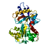
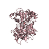
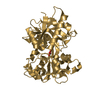
| Entry | Database: PDB / ID: 1jqf | ||||||
|---|---|---|---|---|---|---|---|
| Title | Human Transferrin N-Lobe Mutant H249Q | ||||||
 Components Components | TRANSFERRIN | ||||||
 Keywords Keywords | TRANSPORT PROTEIN / iron binding protein | ||||||
| Function / homology |  Function and homology information Function and homology informationiron chaperone activity / transferrin receptor binding / Transferrin endocytosis and recycling / basal part of cell / endocytic vesicle / clathrin-coated pit / ferric iron binding / basal plasma membrane / osteoclast differentiation / cellular response to iron ion ...iron chaperone activity / transferrin receptor binding / Transferrin endocytosis and recycling / basal part of cell / endocytic vesicle / clathrin-coated pit / ferric iron binding / basal plasma membrane / osteoclast differentiation / cellular response to iron ion / Post-translational protein phosphorylation / clathrin-coated endocytic vesicle membrane / iron ion transport / Iron uptake and transport / ferrous iron binding / regulation of iron ion transport / HFE-transferrin receptor complex / recycling endosome / regulation of protein stability / positive regulation of receptor-mediated endocytosis / multicellular organismal-level iron ion homeostasis / Regulation of Insulin-like Growth Factor (IGF) transport and uptake by Insulin-like Growth Factor Binding Proteins (IGFBPs) / antibacterial humoral response / late endosome / positive regulation of proteasomal ubiquitin-dependent protein catabolic process / Platelet degranulation / Cargo recognition for clathrin-mediated endocytosis / Clathrin-mediated endocytosis / cytoplasmic vesicle / secretory granule lumen / blood microparticle / vesicle / transmembrane transporter binding / intracellular iron ion homeostasis / early endosome / endosome membrane / apical plasma membrane / endoplasmic reticulum lumen / perinuclear region of cytoplasm / enzyme binding / cell surface / extracellular space / extracellular exosome / extracellular region / plasma membrane Similarity search - Function | ||||||
| Biological species |  Homo sapiens (human) Homo sapiens (human) | ||||||
| Method |  X-RAY DIFFRACTION / MOLECULAR REPLACEMENT / Resolution: 1.85 Å X-RAY DIFFRACTION / MOLECULAR REPLACEMENT / Resolution: 1.85 Å | ||||||
 Authors Authors | Baker, H.M. / Mason, A.B. / He, Q.-Y. / MacGillivray, R.T.A. / Baker, E.N. | ||||||
 Citation Citation | Journal: Biochemistry / Year: 2001 Title: Ligand variation in the transferrin family: the crystal structure of the H249Q mutant of the human transferrin N-lobe as a model for iron binding in insect transferrins. Authors: Baker, H.M. / Mason, A.B. / He, Q.Y. / MacGillivray, R.T. / Baker, E.N. | ||||||
| History |
|
- Structure visualization
| Structure viewer | Molecule: MolmilJmol/JSmol |
|---|
- Downloads & links
-Download
| PDBx/mmCIF format | 1jqf.cif.gz | 79.6 KB | Display | PDBx/mmCIF format |
|---|---|---|---|---|
| PDB format | pdb1jqf.ent.gz | 59.2 KB | Display | PDB format |
| PDBx/mmJSON format | 1jqf.json.gz | Tree view | PDBx/mmJSON format | |
| Others |  Other downloads Other downloads |
-Validation report
| Summary document | 1jqf_validation.pdf.gz | 429 KB | Display | wwPDB validaton report |
|---|---|---|---|---|
| Full document | 1jqf_full_validation.pdf.gz | 431.7 KB | Display | |
| Data in XML | 1jqf_validation.xml.gz | 15.6 KB | Display | |
| Data in CIF | 1jqf_validation.cif.gz | 22.2 KB | Display | |
| Arichive directory | https://data.pdbj.org/pub/pdb/validation_reports/jq/1jqfftp://data.pdbj.org/pub/pdb/validation_reports/jq/1jqf | HTTPS FTP |
-Related structure data
| Related structure data | 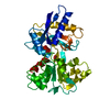 1a8eS S: Starting model for refinement |
|---|---|
| Similar structure data |


-Links
 PDBj
PDBj


- Assembly
| Deposited unit | 
| ||||||||
|---|---|---|---|---|---|---|---|---|---|
| 1 |
| ||||||||
| Unit cell |
|
-Components
| #1: Protein | Mass: 36891.945 Da / Num. of mol.: 1 / Fragment: N-TERMINUS (Residues 20-353) / Mutation: H249Q Source method: isolated from a genetically manipulated source Source: (gene. exp.) Homo sapiens (human) / Plasmid: pNUT / Cell (production host): Kidney / Production host:   Cricetulus griseus (Chinese hamster) / References: UniProt: P02787 Cricetulus griseus (Chinese hamster) / References: UniProt: P02787 |
|---|---|
| #2: Chemical | ChemComp-CO3 /   Mass: 60.009 Da / Num. of mol.: 1 / Source method: obtained synthetically / Formula: CO3 Mass: 60.009 Da / Num. of mol.: 1 / Source method: obtained synthetically / Formula: CO3 |
| #3: Chemical | ChemComp-FE /   Mass: 55.845 Da / Num. of mol.: 1 / Source method: obtained synthetically / Formula: Fe Mass: 55.845 Da / Num. of mol.: 1 / Source method: obtained synthetically / Formula: Fe |
| #4: Chemical | ChemComp-K /   Mass: 39.098 Da / Num. of mol.: 1 / Source method: obtained synthetically / Formula: K Mass: 39.098 Da / Num. of mol.: 1 / Source method: obtained synthetically / Formula: K |
| #5: Water | ChemComp-HOH /  Mass: 18.015 Da / Num. of mol.: 192 / Source method: isolated from a natural source / Formula: H2O Mass: 18.015 Da / Num. of mol.: 192 / Source method: isolated from a natural source / Formula: H2O |
| Has protein modification | Y |
-Experimental details
-Experiment
| Experiment | Method: X-RAY DIFFRACTION / Number of used crystals: 1 |
|---|
- Sample preparation
| Crystal | Density Matthews: 2.3 Å3/Da / Density % sol: 47 % | ||||||||||||||||||||
|---|---|---|---|---|---|---|---|---|---|---|---|---|---|---|---|---|---|---|---|---|---|
| Crystal grow | Temperature: 291 K / Method: vapor diffusion, hanging drop / pH: 7.4 Details: Hanging drop. Protein solution 35 mg/ml protein in 0.1M ammonium bicarbonate pH 7.4. Reservoir solution 0.1M potassium acetate, pH 7.4, 20-25% PEG 3350. Equal volumes of protein and ...Details: Hanging drop. Protein solution 35 mg/ml protein in 0.1M ammonium bicarbonate pH 7.4. Reservoir solution 0.1M potassium acetate, pH 7.4, 20-25% PEG 3350. Equal volumes of protein and reservoir solutions mixed., VAPOR DIFFUSION, HANGING DROP, temperature 291K | ||||||||||||||||||||
| Crystal grow | *PLUS Details: used microseeding | ||||||||||||||||||||
| Components of the solutions | *PLUS
|
-Data collection
| Diffraction | Mean temperature: 110 K |
|---|---|
| Diffraction source | Source: ROTATING ANODE / Type: RIGAKU RU300 / Wavelength: 1.54 Å |
| Detector | Type: MARRESEARCH / Detector: IMAGE PLATE / Date: Oct 1, 2000 / Details: mirrors |
| Radiation | Monochromator: TR mirrors / Protocol: SINGLE WAVELENGTH / Monochromatic (M) / Laue (L): M / Scattering type: x-ray |
| Radiation wavelength | Wavelength: 1.54 Å / Relative weight: 1 |
| Reflection | Resolution: 1.85→30 Å / Num. all: 30081 / % possible obs: 99.4 % / Redundancy: 4.6 % / Rmerge(I) obs: 0.087 / Net I/σ(I): 17 |
| Reflection shell | Resolution: 1.85→1.92 Å / Redundancy: 3.2 % / Rmerge(I) obs: 0.394 / Mean I/σ(I) obs: 2.9 / Num. unique all: 2847 / % possible all: 96.5 |
| Reflection | *PLUS Lowest resolution: 30 Å / Num. obs: 30081 |
| Reflection shell | *PLUS % possible obs: 96.5 % |
- Processing
| Software |
| ||||||||||||||||
|---|---|---|---|---|---|---|---|---|---|---|---|---|---|---|---|---|---|
| Refinement | Method to determine structure: MOLECULAR REPLACEMENT Starting model: PDB entry 1A8E Resolution: 1.85→30 Å / Isotropic thermal model: isotropic / Cross valid method: THROUGHOUT / Stereochemistry target values: Engh & Huber
| ||||||||||||||||
| Refinement step | Cycle: LAST / Resolution: 1.85→30 Å
| ||||||||||||||||
| Refine LS restraints |
| ||||||||||||||||
| Software | *PLUS Name: CNS / Classification: refinement | ||||||||||||||||
| Refinement | *PLUS Lowest resolution: 30 Å / Rfactor obs: 0.221 | ||||||||||||||||
| Solvent computation | *PLUS | ||||||||||||||||
| Displacement parameters | *PLUS |
