 Movie
Movie Controller

[English] 日本語
 Yorodumi
- PDB-1jku: Crystal Structure of Manganese Catalase from Lactobacillus plantarum -
+ Open data

- Basic information
| Entry | Database: PDB / ID: 1jku | ||||||
|---|---|---|---|---|---|---|---|
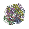
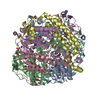
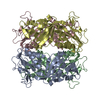
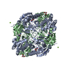
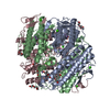
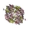
| Title | Crystal Structure of Manganese Catalase from Lactobacillus plantarum | ||||||
 Components Components | pseudocatalase | ||||||
 Keywords Keywords | OXIDOREDUCTASE / hexamer / catalase dimanganese / metalloenzyme | ||||||
| Function / homology |  Function and homology information Function and homology information | ||||||
| Biological species |  Lactobacillus plantarum (bacteria) Lactobacillus plantarum (bacteria) | ||||||
| Method |  X-RAY DIFFRACTION / SYNCHROTRON / MOLECULAR REPLACEMENT / Resolution: 1.84 Å X-RAY DIFFRACTION / SYNCHROTRON / MOLECULAR REPLACEMENT / Resolution: 1.84 Å | ||||||
 Authors Authors | Barynin, V.V. / Whittaker, M.M. / Antonyuk, S.V. / Lamzin, V.S. / Harrison, P.M. / Artymiuk, P.J. / Whittaker, J.W. | ||||||
 Citation Citation | Journal: Structure / Year: 2001 Title: Crystal structure of manganese catalase from Lactobacillus plantarum. Authors: Barynin, V.V. / Whittaker, M.M. / Antonyuk, S.V. / Lamzin, V.S. / Harrison, P.M. / Artymiuk, P.J. / Whittaker, J.W. | ||||||
| History |
|
- Structure visualization
| Structure viewer | Molecule: MolmilJmol/JSmol |
|---|
- Downloads & links
-Download
| PDBx/mmCIF format | 1jku.cif.gz | 338.7 KB | Display | PDBx/mmCIF format |
|---|---|---|---|---|
| PDB format | pdb1jku.ent.gz | 272.5 KB | Display | PDB format |
| PDBx/mmJSON format | 1jku.json.gz | Tree view | PDBx/mmJSON format | |
| Others |  Other downloads Other downloads |
-Validation report
| Summary document | 1jku_validation.pdf.gz | 408.4 KB | Display | wwPDB validaton report |
|---|---|---|---|---|
| Full document | 1jku_full_validation.pdf.gz | 445 KB | Display | |
| Data in XML | 1jku_validation.xml.gz | 35.8 KB | Display | |
| Data in CIF | 1jku_validation.cif.gz | 58.3 KB | Display | |
| Arichive directory | https://data.pdbj.org/pub/pdb/validation_reports/jk/1jkuftp://data.pdbj.org/pub/pdb/validation_reports/jk/1jku | HTTPS FTP |
-Related structure data


-Links
 PDBj
PDBj


- Assembly
| Deposited unit | 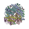
| ||||||||||
|---|---|---|---|---|---|---|---|---|---|---|---|
| 1 |
| ||||||||||
| Unit cell |
|
-Components
| #1: Protein | Mass: 29779.379 Da / Num. of mol.: 6 / Source method: isolated from a natural source / Source: (natural) Lactobacillus plantarum (bacteria) / Strain: ATCC 14431 / References: UniProt: P60355, catalase#2: Chemical | ChemComp-CA /   Mass: 40.078 Da / Num. of mol.: 6 / Source method: obtained synthetically / Formula: Ca Mass: 40.078 Da / Num. of mol.: 6 / Source method: obtained synthetically / Formula: Ca#3: Chemical | ChemComp-OH /   Mass: 17.007 Da / Num. of mol.: 18 / Source method: obtained synthetically / Formula: HO Mass: 17.007 Da / Num. of mol.: 18 / Source method: obtained synthetically / Formula: HO#4: Chemical | ChemComp-MN3 /   Mass: 54.938 Da / Num. of mol.: 12 / Source method: obtained synthetically / Formula: Mn Mass: 54.938 Da / Num. of mol.: 12 / Source method: obtained synthetically / Formula: Mn#5: Water | ChemComp-HOH / |  Mass: 18.015 Da / Num. of mol.: 989 / Source method: isolated from a natural source / Formula: H2O Mass: 18.015 Da / Num. of mol.: 989 / Source method: isolated from a natural source / Formula: H2O |
|---|
-Experimental details
-Experiment
| Experiment | Method: X-RAY DIFFRACTION / Number of used crystals: 1 |
|---|
- Sample preparation
| Crystal | Density Matthews: 2.06 Å3/Da / Density % sol: 40.22 % | ||||||||||||||||||||||||
|---|---|---|---|---|---|---|---|---|---|---|---|---|---|---|---|---|---|---|---|---|---|---|---|---|---|
| Crystal grow | Temperature: 291 K / Method: vapor diffusion, hanging drop / pH: 8.5 Details: PEG 8000, TAPS, pH 8.5, VAPOR DIFFUSION, HANGING DROP at 291K | ||||||||||||||||||||||||
| Crystal grow | *PLUS Temperature: 17 ℃ / pH: 8.7 | ||||||||||||||||||||||||
| Components of the solutions | *PLUS
|
-Data collection
| Diffraction | Mean temperature: 298 K |
|---|---|
| Diffraction source | Source: SYNCHROTRON / Site: EMBL/DESY, HAMBURG  / Beamline: BW7B / Wavelength: 0.89 Å / Beamline: BW7B / Wavelength: 0.89 Å |
| Detector | Type: MARRESEARCH / Detector: IMAGE PLATE / Date: Jun 12, 1996 |
| Radiation | Protocol: SINGLE WAVELENGTH / Monochromatic (M) / Laue (L): M / Scattering type: x-ray |
| Radiation wavelength | Wavelength: 0.89 Å / Relative weight: 1 |
| Reflection | Resolution: 1.84→17 Å / Observed criterion σ(I): -3 |
| Reflection shell | Resolution: 1.839→1.886 Å / Rmerge(I) obs: 0.136 / Mean I/σ(I) obs: 3 / % possible all: 63.6 |
| Reflection | *PLUS Num. obs: 113270 / % possible obs: 90.4 % / Num. measured all: 157987 / Rmerge(I) obs: 0.055 |
| Reflection shell | *PLUS Highest resolution: 1.84 Å / Lowest resolution: 1.87 Å / % possible obs: 63.6 % / Rmerge(I) obs: 0.136 |
- Processing
| Software |
| |||||||||||||||||||||||||||||||||||
|---|---|---|---|---|---|---|---|---|---|---|---|---|---|---|---|---|---|---|---|---|---|---|---|---|---|---|---|---|---|---|---|---|---|---|---|---|
| Refinement | Method to determine structure: MOLECULAR REPLACEMENT Starting model: hexamer of Thermus thermophilus Mn catalase Resolution: 1.84→14.94 Å / Isotropic thermal model: isotropic / Cross valid method: THROUGHOUT / σ(F): 2 / σ(I): 1 / Stereochemistry target values: Engh & Huber
| |||||||||||||||||||||||||||||||||||
| Solvent computation | Solvent model: BABINET MODEL WITH MASK | |||||||||||||||||||||||||||||||||||
| Displacement parameters | Biso mean: 19.072 Å2
| |||||||||||||||||||||||||||||||||||
| Refinement step | Cycle: LAST / Resolution: 1.84→14.94 Å
| |||||||||||||||||||||||||||||||||||
| Refine LS restraints |
| |||||||||||||||||||||||||||||||||||
| LS refinement shell | Resolution: 1.839→1.886 Å / Rfactor Rfree error: 0.128 / Total num. of bins used: 20
| |||||||||||||||||||||||||||||||||||
| Refinement | *PLUS Lowest resolution: 14.9 Å / % reflection Rfree: 5 % / Rfactor all: 0.1469 / Rfactor obs: 0.14691 / Rfactor Rfree: 0.2 / Rfactor Rwork: 0.146 | |||||||||||||||||||||||||||||||||||
| Solvent computation | *PLUS | |||||||||||||||||||||||||||||||||||
| Displacement parameters | *PLUS | |||||||||||||||||||||||||||||||||||
| Refine LS restraints | *PLUS
| |||||||||||||||||||||||||||||||||||
| LS refinement shell | *PLUS Rfactor Rfree: 0.245 / Rfactor Rwork: 0.183 |
