 Movie
Movie Controller

[English] 日本語
 Yorodumi
- PDB-1o9i: Crystal structure of the Y42F mutant of manganese catalase from L... -
+ Open data

- Basic information
| Entry | Database: PDB / ID: 1o9i | ||||||
|---|---|---|---|---|---|---|---|
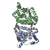
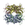
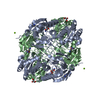
| Title | Crystal structure of the Y42F mutant of manganese catalase from Lactobacillus plantarum at 1.33A resolution | ||||||
 Components Components | Manganese catalase | ||||||
 Keywords Keywords | OXIDOREDUCTASE / HEXAMER / DIMANGANESE CATALASE / METALLOENZYME / PREOXIDASE | ||||||
| Function / homology |  Function and homology information Function and homology information | ||||||
| Biological species |  Lactobacillus plantarum (bacteria) Lactobacillus plantarum (bacteria) | ||||||
| Method |  X-RAY DIFFRACTION / SYNCHROTRON / MOLECULAR REPLACEMENT / Resolution: 1.33 Å X-RAY DIFFRACTION / SYNCHROTRON / MOLECULAR REPLACEMENT / Resolution: 1.33 Å | ||||||
 Authors Authors | Barynin, V.V. / Whittaker, M.M. / Whittaker, J.W. | ||||||
 Citation Citation | Journal: Eur.J.Biochem. / Year: 2003 Title: Outer Sphere Mutagenesis of Lactobacillus Plantarum Manganese Catalase Disrupts the Cluster Core. Mechanistic Implications. Authors: Whittaker, M.M. / Barynin, V.V. / Igarashi, T. / Whittaker, J.W. #1: Journal: Structure / Year: 2001Title: Crystal Structure of Manganese Catalase from Lactobacillus Plantarum Authors: Barynin, V.V. / Whittaker, M.M. / Antonyuk, S.V. / Lamzin, V.S. / Harrison, P.M. / Artymiuk, P.J. / Whittaker, J.W. | ||||||
| History |
|
- Structure visualization
| Structure viewer | Molecule: MolmilJmol/JSmol |
|---|
- Downloads & links
-Download
| PDBx/mmCIF format | 1o9i.cif.gz | 675.8 KB | Display | PDBx/mmCIF format |
|---|---|---|---|---|
| PDB format | pdb1o9i.ent.gz | 558.4 KB | Display | PDB format |
| PDBx/mmJSON format | 1o9i.json.gz | Tree view | PDBx/mmJSON format | |
| Others |  Other downloads Other downloads |
-Validation report
| Arichive directory | https://data.pdbj.org/pub/pdb/validation_reports/o9/1o9iftp://data.pdbj.org/pub/pdb/validation_reports/o9/1o9i | HTTPS FTP |
|---|
-Related structure data
| Related structure data |  1jkvS S: Starting model for refinement |
|---|---|
| Similar structure data |


-Links
 PDBj
PDBj


- Assembly
| Deposited unit | 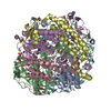
| ||||||||
|---|---|---|---|---|---|---|---|---|---|
| 1 |
| ||||||||
| Unit cell |
|
-Components
-Protein , 1 types, 6 molecules ABCDEF
| #1: Protein | Mass: 29763.379 Da / Num. of mol.: 6 / Mutation: YES Source method: isolated from a genetically manipulated source Source: (gene. exp.) Lactobacillus plantarum (bacteria) / Plasmid: PVMG36PSLPA-LPC / Production host: LACTOBACILLUS PLANTARUM (bacteria) / Strain (production host): KAT- L.PLANTARUM (NCDO 1193) / References: UniProt: P60355, catalase |
|---|
-Non-polymers , 6 types, 1490 molecules 


| #2: Chemical | ChemComp-MN3 /  Mass: 54.938 Da / Num. of mol.: 12 / Source method: obtained synthetically / Formula: Mn Mass: 54.938 Da / Num. of mol.: 12 / Source method: obtained synthetically / Formula: Mn#3: Chemical | ChemComp-CA /  Mass: 40.078 Da / Num. of mol.: 6 / Source method: obtained synthetically / Formula: Ca Mass: 40.078 Da / Num. of mol.: 6 / Source method: obtained synthetically / Formula: Ca#4: Chemical | ChemComp-O /  Mass: 15.999 Da / Num. of mol.: 6 / Source method: obtained synthetically / Formula: O Mass: 15.999 Da / Num. of mol.: 6 / Source method: obtained synthetically / Formula: O#5: Chemical | ChemComp-MES /  Mass: 195.237 Da / Num. of mol.: 5 / Source method: obtained synthetically / Formula: C6H13NO4S / Comment: pH buffer*YM Mass: 195.237 Da / Num. of mol.: 5 / Source method: obtained synthetically / Formula: C6H13NO4S / Comment: pH buffer*YM#6: Chemical |  Mass: 22.990 Da / Num. of mol.: 2 / Source method: obtained synthetically / Formula: Na Mass: 22.990 Da / Num. of mol.: 2 / Source method: obtained synthetically / Formula: Na#7: Water | ChemComp-HOH / | Mass: 18.015 Da / Num. of mol.: 1459 / Source method: isolated from a natural source / Formula: H2O |
|---|
-Details
| Compound details | ENGINEERED |
|---|
-Experimental details
-Experiment
| Experiment | Method: X-RAY DIFFRACTION / Number of used crystals: 1 |
|---|
- Sample preparation
| Crystal | Density Matthews: 1.97 Å3/Da / Density % sol: 37.66 % | ||||||||||||||||||
|---|---|---|---|---|---|---|---|---|---|---|---|---|---|---|---|---|---|---|---|
| Crystal grow | Temperature: 291 K / Method: vapor diffusion, hanging drop / pH: 5.5 Details: PEG 8000, TAPS, PH 8.5, VAPOR DIFFUSION, HANGING DROP AT 291K | ||||||||||||||||||
| Crystal grow | *PLUS Temperature: 18 ℃ / pH: 8.7 / Method: vapor diffusion | ||||||||||||||||||
| Components of the solutions | *PLUS
|
-Data collection
| Diffraction | Mean temperature: 100 K |
|---|---|
| Diffraction source | Source: SYNCHROTRON / Site: SRS  / Beamline: PX9.6 / Wavelength: 0.87 / Beamline: PX9.6 / Wavelength: 0.87 |
| Detector | Type: ADSC CCD / Detector: CCD / Date: May 9, 2001 |
| Radiation | Protocol: SINGLE WAVELENGTH / Monochromatic (M) / Laue (L): M / Scattering type: x-ray |
| Radiation wavelength | Wavelength: 0.87 Å / Relative weight: 1 |
| Reflection | Resolution: 1.33→50 Å / Num. obs: 282601 / % possible obs: 86.7 % / Observed criterion σ(I): -3 / Redundancy: 2.1 % / Biso Wilson estimate: 14.28 Å2 / Rsym value: 0.041 / Net I/σ(I): 14.9 |
| Reflection shell | Resolution: 1.33→1.35 Å / Redundancy: 1.88 % / Mean I/σ(I) obs: 1.7 / Rsym value: 0.34 / % possible all: 80.7 |
| Reflection | *PLUS Highest resolution: 1.33 Å / Lowest resolution: 23 Å / Num. obs: 274725 / Redundancy: 2.14 % / Num. measured all: 588079 / Rmerge(I) obs: 0.041 |
| Reflection shell | *PLUS % possible obs: 80.7 % / Rmerge(I) obs: 0.34 / Mean I/σ(I) obs: 1.7 |
- Processing
| Software |
| ||||||||||||||||||||
|---|---|---|---|---|---|---|---|---|---|---|---|---|---|---|---|---|---|---|---|---|---|
| Refinement | Method to determine structure: MOLECULAR REPLACEMENT Starting model: PDB ENTRY 1JKV Resolution: 1.33→50 Å / SU B: 2.092 / SU ML: 0.043 / σ(F): 0 / ESU R: 0.052 / ESU R Free: 0.047 / Details: HYDROGENS HAVE BEEN ADDED IN THE RIDING POSITIONS
| ||||||||||||||||||||
| Displacement parameters | Biso mean: 16.33 Å2
| ||||||||||||||||||||
| Refinement step | Cycle: LAST / Resolution: 1.33→50 Å
| ||||||||||||||||||||
| Refinement | *PLUS Lowest resolution: 48 Å / % reflection Rfree: 5 % | ||||||||||||||||||||
| Solvent computation | *PLUS | ||||||||||||||||||||
| Displacement parameters | *PLUS | ||||||||||||||||||||
| Refine LS restraints | *PLUS
|
