 Movie
Movie Controller

[English] 日本語
 Yorodumi
- PDB-1jkv: Crystal Structure of Manganese Catalase from Lactobacillus planta... -
+ Open data

- Basic information
| Entry | Database: PDB / ID: 1jkv | ||||||
|---|---|---|---|---|---|---|---|
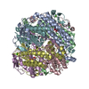
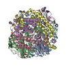
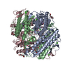
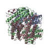
| Title | Crystal Structure of Manganese Catalase from Lactobacillus plantarum complexed with azide | ||||||
 Components Components | pseudocatalase | ||||||
 Keywords Keywords | OXIDOREDUCTASE / hexamer / catalase dimanganese / metalloenzyme | ||||||
| Function / homology |  Function and homology information Function and homology information | ||||||
| Biological species |  Lactobacillus plantarum (bacteria) Lactobacillus plantarum (bacteria) | ||||||
| Method |  X-RAY DIFFRACTION / SYNCHROTRON / MOLECULAR REPLACEMENT / Resolution: 1.39 Å X-RAY DIFFRACTION / SYNCHROTRON / MOLECULAR REPLACEMENT / Resolution: 1.39 Å | ||||||
 Authors Authors | Barynin, V.V. / Whittaker, M.M. / Antonyuk, S.V. / Lamzin, V.S. / Harrison, P.M. / Artymiuk, P.J. / Whittaker, J.W. | ||||||
 Citation Citation | Journal: Structure / Year: 2001 Title: Crystal structure of manganese catalase from Lactobacillus plantarum. Authors: Barynin, V.V. / Whittaker, M.M. / Antonyuk, S.V. / Lamzin, V.S. / Harrison, P.M. / Artymiuk, P.J. / Whittaker, J.W. | ||||||
| History |
|
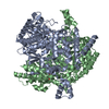
- Structure visualization
| Structure viewer | Molecule: MolmilJmol/JSmol |
|---|
- Downloads & links
-Download
| PDBx/mmCIF format | 1jkv.cif.gz | 682.7 KB | Display | PDBx/mmCIF format |
|---|---|---|---|---|
| PDB format | pdb1jkv.ent.gz | 558.1 KB | Display | PDB format |
| PDBx/mmJSON format | 1jkv.json.gz | Tree view | PDBx/mmJSON format | |
| Others |  Other downloads Other downloads |
-Validation report
| Arichive directory | https://data.pdbj.org/pub/pdb/validation_reports/jk/1jkvftp://data.pdbj.org/pub/pdb/validation_reports/jk/1jkv | HTTPS FTP |
|---|
-Related structure data
| Related structure data |  1jkuSC S: Starting model for refinement C: citing same article ( |
|---|---|
| Similar structure data |

-Links
 PDBj
PDBj


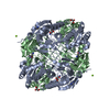
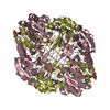
- Assembly
| Deposited unit | 
| ||||||||||
|---|---|---|---|---|---|---|---|---|---|---|---|
| 1 |
| ||||||||||
| Unit cell |
|
-Components
-Protein , 1 types, 6 molecules ABCDEF
| #1: Protein | Mass: 29779.379 Da / Num. of mol.: 6 / Source method: isolated from a natural source / Source: (natural) Lactobacillus plantarum (bacteria) / Strain: ATCC 14431 / References: UniProt: P60355, catalase |
|---|
-Non-polymers , 6 types, 1591 molecules 


| #2: Chemical | ChemComp-CA /  Mass: 40.078 Da / Num. of mol.: 6 / Source method: obtained synthetically / Formula: Ca Mass: 40.078 Da / Num. of mol.: 6 / Source method: obtained synthetically / Formula: Ca#3: Chemical | ChemComp-OH /  Mass: 17.007 Da / Num. of mol.: 18 / Source method: obtained synthetically / Formula: HO Mass: 17.007 Da / Num. of mol.: 18 / Source method: obtained synthetically / Formula: HO#4: Chemical | ChemComp-AZI /  Mass: 42.020 Da / Num. of mol.: 6 / Source method: obtained synthetically / Formula: N3 Mass: 42.020 Da / Num. of mol.: 6 / Source method: obtained synthetically / Formula: N3#5: Chemical | ChemComp-MN3 /  Mass: 54.938 Da / Num. of mol.: 12 / Source method: obtained synthetically / Formula: Mn Mass: 54.938 Da / Num. of mol.: 12 / Source method: obtained synthetically / Formula: Mn#6: Chemical | ChemComp-EDO /  Mass: 62.068 Da / Num. of mol.: 15 / Source method: obtained synthetically / Formula: C2H6O2 Mass: 62.068 Da / Num. of mol.: 15 / Source method: obtained synthetically / Formula: C2H6O2#7: Water | ChemComp-HOH / | Mass: 18.015 Da / Num. of mol.: 1534 / Source method: isolated from a natural source / Formula: H2O |
|---|
-Experimental details
-Experiment
| Experiment | Method: X-RAY DIFFRACTION / Number of used crystals: 1 |
|---|
- Sample preparation
| Crystal | Density Matthews: 2.02 Å3/Da / Density % sol: 39.15 % | ||||||||||||||||||||||||
|---|---|---|---|---|---|---|---|---|---|---|---|---|---|---|---|---|---|---|---|---|---|---|---|---|---|
| Crystal grow | Temperature: 291 K / Method: vapor diffusion, hanging drop / pH: 8.7 Details: PEG 8000, TAPS, pH 8.5, VAPOR DIFFUSION, HANGING DROP at 291K, pH 8.7 | ||||||||||||||||||||||||
| Crystal grow | *PLUS Temperature: 17 ℃ | ||||||||||||||||||||||||
| Components of the solutions | *PLUS
|
-Data collection
| Diffraction | Mean temperature: 100 K |
|---|---|
| Diffraction source | Source: SYNCHROTRON / Site: SRS  / Beamline: PX9.6 / Wavelength: 0.811 Å / Beamline: PX9.6 / Wavelength: 0.811 Å |
| Detector | Type: ADSC QUANTUM 4 / Detector: CCD / Date: Sep 17, 1999 |
| Radiation | Protocol: SINGLE WAVELENGTH / Monochromatic (M) / Laue (L): M / Scattering type: x-ray |
| Radiation wavelength | Wavelength: 0.811 Å / Relative weight: 1 |
| Reflection | Resolution: 1.39→17 Å / Num. all: 269006 / Num. obs: 269006 / % possible obs: 95.5 % / Observed criterion σ(I): -3 / Redundancy: 2.5 % / Rmerge(I) obs: 0.05 / Rsym value: 5 / Net I/σ(I): 16.4 |
| Reflection shell | Resolution: 1.39→1.43 Å / Redundancy: 2.1 % / Rmerge(I) obs: 0.14 / Mean I/σ(I) obs: 4.1 / Num. unique all: 15234 / Rsym value: 14 / % possible all: 78.3 |
| Reflection | *PLUS Highest resolution: 1.4 Å / Num. measured all: 663270 / Rmerge(I) obs: 0.05 |
| Reflection shell | *PLUS Highest resolution: 1.4 Å / Lowest resolution: 1.42 Å / % possible obs: 78.3 % / Rmerge(I) obs: 0.14 / Mean I/σ(I) obs: 4.13 |
- Processing
| Software |
| |||||||||||||||||||||||||||||||||||
|---|---|---|---|---|---|---|---|---|---|---|---|---|---|---|---|---|---|---|---|---|---|---|---|---|---|---|---|---|---|---|---|---|---|---|---|---|
| Refinement | Method to determine structure: MOLECULAR REPLACEMENT Starting model: PDB ENTRY 1JKU Resolution: 1.39→17 Å / Isotropic thermal model: anisotropic / Cross valid method: THROUGHOUT / σ(F): 2 / σ(I): 1 / Stereochemistry target values: Engh & Huber
| |||||||||||||||||||||||||||||||||||
| Solvent computation | Solvent model: BABINET MODEL WITH MASK | |||||||||||||||||||||||||||||||||||
| Displacement parameters | Biso mean: 11.975 Å2
| |||||||||||||||||||||||||||||||||||
| Refinement step | Cycle: LAST / Resolution: 1.39→17 Å
| |||||||||||||||||||||||||||||||||||
| Refine LS restraints |
| |||||||||||||||||||||||||||||||||||
| LS refinement shell | Resolution: 1.39→1.43 Å / Rfactor Rfree error: 0.043 / Total num. of bins used: 20
| |||||||||||||||||||||||||||||||||||
| Refinement | *PLUS Highest resolution: 1.4 Å / Lowest resolution: 17 Å / Num. reflection Rfree: 13288 / % reflection Rfree: 5 % / Rfactor all: 0.1037 / Rfactor obs: 0.10509 / Rfactor Rfree: 0.13 / Rfactor Rwork: 0.104 | |||||||||||||||||||||||||||||||||||
| Solvent computation | *PLUS | |||||||||||||||||||||||||||||||||||
| Displacement parameters | *PLUS | |||||||||||||||||||||||||||||||||||
| Refine LS restraints | *PLUS
| |||||||||||||||||||||||||||||||||||
| LS refinement shell | *PLUS Rfactor Rfree: 0.172 / Rfactor Rwork: 0.119 |
