 Movie
Movie Controller


+ Open data

- Basic information
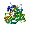
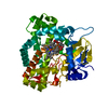
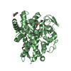
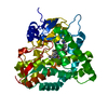
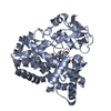
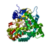
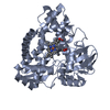
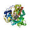
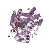
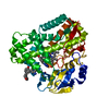
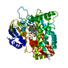
| Entry | Database: PDB / ID: 1jio | ||||||
|---|---|---|---|---|---|---|---|
| Title | P450eryF/6DEB | ||||||
 Components Components | CYTOCHROME P450 107A1 | ||||||
 Keywords Keywords | HYDROLASE / Cytochrome P450 / P450 / 6-deoxyerythronolide B / 6-DEB | ||||||
| Function / homology |  Function and homology information Function and homology information6-deoxyerythronolide B hydroxylase / erythromycin biosynthetic process / oxidoreductase activity, acting on paired donors, with incorporation or reduction of molecular oxygen / monooxygenase activity / iron ion binding / heme binding / cytoplasm Similarity search - Function | ||||||
| Biological species |  Saccharopolyspora erythraea (bacteria) Saccharopolyspora erythraea (bacteria) | ||||||
| Method |  X-RAY DIFFRACTION / MOLECULAR REPLACEMENT / Resolution: 2.1 Å X-RAY DIFFRACTION / MOLECULAR REPLACEMENT / Resolution: 2.1 Å | ||||||
 Authors Authors | Cupp-Vickery, J.R. / Garcia, C. / Hofacre, A. / McGee-Estrada, K. | ||||||
 Citation Citation | Journal: J.Mol.Biol. / Year: 2001 Title: Ketoconazole-induced conformational changes in the active site of cytochrome P450eryF. Authors: Cupp-Vickery, J.R. / Garcia, C. / Hofacre, A. / McGee-Estrada, K. | ||||||
| History |
|
- Structure visualization
| Structure viewer | Molecule: MolmilJmol/JSmol |
|---|
- Downloads & links
-Download
| PDBx/mmCIF format | 1jio.cif.gz | 96 KB | Display | PDBx/mmCIF format |
|---|---|---|---|---|
| PDB format | pdb1jio.ent.gz | 71 KB | Display | PDB format |
| PDBx/mmJSON format | 1jio.json.gz | Tree view | PDBx/mmJSON format | |
| Others |  Other downloads Other downloads |
-Validation report
| Summary document | 1jio_validation.pdf.gz | 1.2 MB | Display | wwPDB validaton report |
|---|---|---|---|---|
| Full document | 1jio_full_validation.pdf.gz | 1.2 MB | Display | |
| Data in XML | 1jio_validation.xml.gz | 18.8 KB | Display | |
| Data in CIF | 1jio_validation.cif.gz | 25.8 KB | Display | |
| Arichive directory | https://data.pdbj.org/pub/pdb/validation_reports/ji/1jioftp://data.pdbj.org/pub/pdb/validation_reports/ji/1jio | HTTPS FTP |
-Related structure data

-Links
 PDBj
PDBj
- Assembly
| Deposited unit | 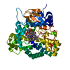
| ||||||||
|---|---|---|---|---|---|---|---|---|---|
| 1 |
| ||||||||
| Unit cell |
|
-Components
| #1: Protein | Mass: 45021.855 Da / Num. of mol.: 1 Source method: isolated from a genetically manipulated source Source: (gene. exp.) Saccharopolyspora erythraea (bacteria) / Plasmid: pTrc99A / Production host: |
|---|---|
| #2: Chemical | ChemComp-HEM /   Mass: 616.487 Da / Num. of mol.: 1 / Source method: obtained synthetically / Formula: C34H32FeN4O4 Mass: 616.487 Da / Num. of mol.: 1 / Source method: obtained synthetically / Formula: C34H32FeN4O4 |
| #3: Chemical | ChemComp-DEB / 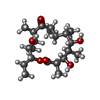  Mass: 386.523 Da / Num. of mol.: 1 / Source method: obtained synthetically / Formula: C21H38O6 Mass: 386.523 Da / Num. of mol.: 1 / Source method: obtained synthetically / Formula: C21H38O6 |
| #4: Water | ChemComp-HOH /  Mass: 18.015 Da / Num. of mol.: 120 / Source method: isolated from a natural source / Formula: H2O Mass: 18.015 Da / Num. of mol.: 120 / Source method: isolated from a natural source / Formula: H2O |
-Experimental details
-Experiment
| Experiment | Method: X-RAY DIFFRACTION / Number of used crystals: 1 |
|---|
- Sample preparation
| Crystal | Density Matthews: 2.33 Å3/Da / Density % sol: 47.28 % | ||||||||||||||||||||||||||||||
|---|---|---|---|---|---|---|---|---|---|---|---|---|---|---|---|---|---|---|---|---|---|---|---|---|---|---|---|---|---|---|---|
| Crystal grow | Temperature: 298 K / Method: vapor diffusion, hanging drop / pH: 8.5 Details: peg 4000, pH 8.5, VAPOR DIFFUSION, HANGING DROP, temperature 298K | ||||||||||||||||||||||||||||||
| Crystal grow | *PLUS | ||||||||||||||||||||||||||||||
| Components of the solutions | *PLUS
|
-Data collection
| Diffraction | Mean temperature: 170 K |
|---|---|
| Diffraction source | Source: ROTATING ANODE / Type: RIGAKU / Wavelength: 1.54 Å |
| Detector | Type: RIGAKU RAXIS / Detector: IMAGE PLATE / Date: Jan 1, 2000 |
| Radiation | Protocol: SINGLE WAVELENGTH / Monochromatic (M) / Laue (L): M / Scattering type: x-ray |
| Radiation wavelength | Wavelength: 1.54 Å / Relative weight: 1 |
| Reflection | Resolution: 2.1→50 Å / Num. all: 25292 / Num. obs: 23472 / % possible obs: 92.8 % / Observed criterion σ(F): 1 |
| Reflection | *PLUS Highest resolution: 2.3 Å / Lowest resolution: 40 Å / Num. obs: 19170 / % possible obs: 93.8 % / Num. measured all: 128630 / Rmerge(I) obs: 0.061 |
| Reflection shell | *PLUS % possible obs: 94.4 % / Mean I/σ(I) obs: 2 |
- Processing
| Software |
| |||||||||||||||||||||||||
|---|---|---|---|---|---|---|---|---|---|---|---|---|---|---|---|---|---|---|---|---|---|---|---|---|---|---|
| Refinement | Method to determine structure: MOLECULAR REPLACEMENT Starting model: P450eryF without substrate Resolution: 2.1→50 Å / σ(F): 1
| |||||||||||||||||||||||||
| Refinement step | Cycle: LAST / Resolution: 2.1→50 Å
| |||||||||||||||||||||||||
| Refinement | *PLUS Highest resolution: 2.3 Å / Lowest resolution: 40 Å / % reflection Rfree: 10 % / Rfactor obs: 0.184 / Rfactor Rfree: 0.23 / Rfactor Rwork: 0.184 | |||||||||||||||||||||||||
| Solvent computation | *PLUS | |||||||||||||||||||||||||
| Displacement parameters | *PLUS |
