carbohydrate catabolic process / peptidoglycan catabolic process / cell wall macromolecule catabolic process / lysozyme / lysozyme activity / killing of cells of another organism / defense response to bacterium / extracellular region Similarity search - Function
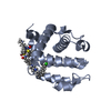
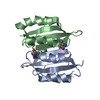
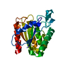
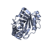
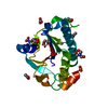
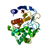
Glycosyl hydrolases family 25, active site / Glycosyl hydrolases family 25 active site signature. / Glycoside hydrolase, family 25 subgroup / Glycosyl hydrolases family 25 / Glycoside hydrolase, family 25 / Glycosyl hydrolases family 25 / Glycosyl hydrolase family 25 (GH25) domain profile. / Glycosidases / Glycoside hydrolase superfamily / TIM Barrel ...Glycosyl hydrolases family 25, active site / Glycosyl hydrolases family 25 active site signature. / Glycoside hydrolase, family 25 subgroup / Glycosyl hydrolases family 25 / Glycoside hydrolase, family 25 / Glycosyl hydrolases family 25 / Glycosyl hydrolase family 25 (GH25) domain profile. / Glycosidases / Glycoside hydrolase superfamily / TIM Barrel / Alpha-Beta Barrel / Alpha Beta Similarity search - Domain/homology
SEQUENCE NO DATABASE REFERENCE SEQUENCE WAS IDENTIFIED FOR 1,4-BETA-N-ACETYLMURAMIDASE M1 OF ... SEQUENCE NO DATABASE REFERENCE SEQUENCE WAS IDENTIFIED FOR 1,4-BETA-N-ACETYLMURAMIDASE M1 OF STREPTOMYCES COELICOLOR. THE DATABASE REFERENCE LISTED IS FOR THE CORRESPONDING PROTEIN IN A CLOSELY RELATED SPECIES OF STREPTOMYCES BACTERIA, STREPTOMYCES GLOBISPORUS.
In the structure databanks used in Yorodumi, some data are registered as the other names, "COVID-19 virus" and "2019-nCoV". Here are the details of the virus and the list of structure data.
Jan 31, 2019. EMDB accession codes are about to change! (news from PDBe EMDB page)
EMDB accession codes are about to change! (news from PDBe EMDB page)
The allocation of 4 digits for EMDB accession codes will soon come to an end. Whilst these codes will remain in use, new EMDB accession codes will include an additional digit and will expand incrementally as the available range of codes is exhausted. The current 4-digit format prefixed with “EMD-” (i.e. EMD-XXXX) will advance to a 5-digit format (i.e. EMD-XXXXX), and so on. It is currently estimated that the 4-digit codes will be depleted around Spring 2019, at which point the 5-digit format will come into force.
The EM Navigator/Yorodumi systems omit the EMD- prefix.
Related info.:Q: What is EMD? / ID/Accession-code notation in Yorodumi/EM Navigator
Yorodumi is a browser for structure data from EMDB, PDB, SASBDB, etc.
This page is also the successor to EM Navigator detail page, and also detail information page/front-end page for Omokage search.
The word "yorodu" (or yorozu) is an old Japanese word meaning "ten thousand". "mi" (miru) is to see.
Related info.:EMDB / PDB / SASBDB / Comparison of 3 databanks / Yorodumi Search / Aug 31, 2016. New EM Navigator & Yorodumi / Yorodumi Papers / Jmol/JSmol / Function and homology information / Changes in new EM Navigator and Yorodumi
 Movie
Movie Controller

 Yorodumi
 Open data
 Basic information
 Components
 Keywords
 Function and homology information
 Streptomyces coelicolor (bacteria)
 X-RAY DIFFRACTION /
  Authors
 Citation
 Structure visualization
 Downloads & links
 Other downloads
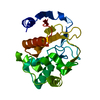

 PDBj
PDBj
 Assembly
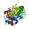

 Mass: 35.453 Da / Num. of mol.: 8 / Source method: obtained synthetically / Formula: Cl
 Mass: 18.015 Da / Num. of mol.: 398 / Source method: isolated from a natural source / Formula: H2O
 Sample preparation
 Processing