 Movie
Movie Controller


+ Open data

- Basic information
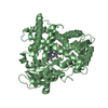
| Entry | Database: PDB / ID: 1jbv | ||||||
|---|---|---|---|---|---|---|---|
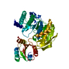
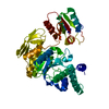
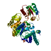
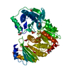
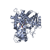
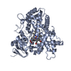
| Title | FPGS-AMPPCP complex | ||||||
 Components Components | FOLYLPOLYGLUTAMATE SYNTHASE | ||||||
 Keywords Keywords | LIGASE / FPGS AMPPCP complex | ||||||
| Function / homology |  Function and homology information Function and homology informationtetrahydrofolate synthase / dihydrofolate synthase activity / tetrahydrofolylpolyglutamate synthase activity / one-carbon metabolic process / ATP binding / metal ion binding / cytoplasm Similarity search - Function | ||||||
| Biological species |  Lactobacillus casei (bacteria) Lactobacillus casei (bacteria) | ||||||
| Method |  X-RAY DIFFRACTION / MOLECULAR REPLACEMENT / Resolution: 1.95 Å X-RAY DIFFRACTION / MOLECULAR REPLACEMENT / Resolution: 1.95 Å | ||||||
 Authors Authors | Sun, X. / Cross, J.A. / Bognar, A.L. / Baker, E.N. / Smith, C.A. | ||||||
 Citation Citation | Journal: J.Mol.Biol. / Year: 2001 Title: Folate-binding triggers the activation of folylpolyglutamate synthetase. Authors: Sun, X. / Cross, J.A. / Bognar, A.L. / Baker, E.N. / Smith, C.A. | ||||||
| History |
|
- Structure visualization
| Structure viewer | Molecule: MolmilJmol/JSmol |
|---|
- Downloads & links
-Download
| PDBx/mmCIF format | 1jbv.cif.gz | 100.4 KB | Display | PDBx/mmCIF format |
|---|---|---|---|---|
| PDB format | pdb1jbv.ent.gz | 73.5 KB | Display | PDB format |
| PDBx/mmJSON format | 1jbv.json.gz | Tree view | PDBx/mmJSON format | |
| Others |  Other downloads Other downloads |
-Validation report
| Arichive directory | https://data.pdbj.org/pub/pdb/validation_reports/jb/1jbvftp://data.pdbj.org/pub/pdb/validation_reports/jb/1jbv | HTTPS FTP |
|---|
-Related structure data
| Related structure data | 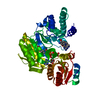 1jbwC 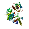 1fgsS S: Starting model for refinement C: citing same article ( |
|---|---|
| Similar structure data |


-Links
 PDBj
PDBj


- Assembly
| Deposited unit | 
| ||||||||
|---|---|---|---|---|---|---|---|---|---|
| 1 |
| ||||||||
| Unit cell |
|
-Components
| #1: Protein | Mass: 46697.016 Da / Num. of mol.: 1 Source method: isolated from a genetically manipulated source Source: (gene. exp.) Lactobacillus casei (bacteria) / Plasmid: pCYB / Species (production host): Escherichia coli / Production host: | ||||
|---|---|---|---|---|---|
| #2: Chemical |   Mass: 24.305 Da / Num. of mol.: 2 / Source method: obtained synthetically / Formula: Mg Mass: 24.305 Da / Num. of mol.: 2 / Source method: obtained synthetically / Formula: Mg#3: Chemical | ChemComp-ACP / |   Mass: 505.208 Da / Num. of mol.: 1 / Source method: obtained synthetically / Formula: C11H18N5O12P3 / Comment: AMP-PCP, energy-carrying molecule analogue*YM Mass: 505.208 Da / Num. of mol.: 1 / Source method: obtained synthetically / Formula: C11H18N5O12P3 / Comment: AMP-PCP, energy-carrying molecule analogue*YM#4: Water | ChemComp-HOH / |  Mass: 18.015 Da / Num. of mol.: 307 / Source method: isolated from a natural source / Formula: H2O Mass: 18.015 Da / Num. of mol.: 307 / Source method: isolated from a natural source / Formula: H2O |
-Experimental details
-Experiment
| Experiment | Method: X-RAY DIFFRACTION / Number of used crystals: 1 |
|---|
- Sample preparation
| Crystal | Density Matthews: 2.06 Å3/Da / Density % sol: 40.31 % | ||||||||||||||||||||||||||||||
|---|---|---|---|---|---|---|---|---|---|---|---|---|---|---|---|---|---|---|---|---|---|---|---|---|---|---|---|---|---|---|---|
| Crystal grow | *PLUS pH: 5.3 / Method: vapor diffusion, hanging dropDetails: Sun, X., (1998) Proc. Natl. Acad. Sci. USA, 95, 6647. | ||||||||||||||||||||||||||||||
| Components of the solutions | *PLUS
|
-Data collection
| Diffraction | Mean temperature: 113 K |
|---|---|
| Diffraction source | Source: ROTATING ANODE / Type: RIGAKU RU300 / Wavelength: 1.5418 Å |
| Detector | Type: MARRESEARCH / Detector: IMAGE PLATE / Date: Jan 10, 2000 / Details: mirrors |
| Radiation | Monochromator: Mar mirrors / Protocol: SINGLE WAVELENGTH / Monochromatic (M) / Laue (L): M / Scattering type: x-ray |
| Radiation wavelength | Wavelength: 1.5418 Å / Relative weight: 1 |
| Reflection | Resolution: 1.9→100 Å / Num. all: 28314 / Num. obs: 28314 / % possible obs: 94 % / Redundancy: 6.9 % / Rmerge(I) obs: 0.051 / Net I/σ(I): 23.4 |
| Reflection shell | Resolution: 1.9→1.97 Å / Rmerge(I) obs: 0.295 / Mean I/σ(I) obs: 3.2 / % possible all: 86.1 |
| Reflection | *PLUS Highest resolution: 1.9 Å / Lowest resolution: 100 Å / % possible obs: 94 % / Num. measured all: 195787 |
| Reflection shell | *PLUS Highest resolution: 1.9 Å / % possible obs: 86.1 % |
- Processing
| Software |
| |||||||||||||||||||||||||
|---|---|---|---|---|---|---|---|---|---|---|---|---|---|---|---|---|---|---|---|---|---|---|---|---|---|---|
| Refinement | Method to determine structure: MOLECULAR REPLACEMENT Starting model: PDB ENTRY 1FGS Resolution: 1.95→100 Å / Cross valid method: THROUGHOUT / Stereochemistry target values: Engh & Huber
| |||||||||||||||||||||||||
| Refinement step | Cycle: LAST / Resolution: 1.95→100 Å
| |||||||||||||||||||||||||
| Refine LS restraints |
| |||||||||||||||||||||||||
| Software | *PLUS Name: CNS / Version: 1 / Classification: refinement | |||||||||||||||||||||||||
| Refinement | *PLUS Lowest resolution: 100 Å / % reflection Rfree: 5 % / Rfactor obs: 0.194 | |||||||||||||||||||||||||
| Solvent computation | *PLUS | |||||||||||||||||||||||||
| Displacement parameters | *PLUS |
