 Movie
Movie Controller

[English] 日本語
 Yorodumi
- PDB-1j8l: Molecular and Crystal Structure of D(CGCAAATTMO4CGCG): the Watson... -
+ Open data

- Basic information
| Entry | Database: PDB / ID: 1j8l | ||||||||||||||||||
|---|---|---|---|---|---|---|---|---|---|---|---|---|---|---|---|---|---|---|---|
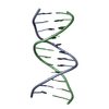
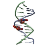
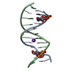
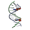
| Title | Molecular and Crystal Structure of D(CGCAAATTMO4CGCG): the Watson-Crick Type N4-Methoxycytidine/Adenosine Base Pair in B-DNA | ||||||||||||||||||
 Components Components | DNA (5'-D(* Keywords KeywordsDNA / N4-METHOXYCYTOSINE / DNA DAMAGE / B-DNA / DOUBLE HELIX / DEOXYRIBONUCLEIC ACID | Function / homology | DNA / DNA (> 10) |  Function and homology information Function and homology informationMethod |  X-RAY DIFFRACTION / SYNCHROTRON / MOLECULAR REPLACEMENT / Resolution: 1.6 Å X-RAY DIFFRACTION / SYNCHROTRON / MOLECULAR REPLACEMENT / Resolution: 1.6 Å  Authors AuthorsHossain, M.T. / Sunami, T. / Tsunoda, M. / Hikima, T. / Chatake, T. / Ueno, Y. / Matsuda, A. / Takenaka, A. |  CitationJournal: Nucleic Acids Res. / Year: 2001 CitationJournal: Nucleic Acids Res. / Year: 2001Title: Crystallographic studies on damaged DNAs IV. N(4)-methoxycytosine shows a second face for Watson-Crick base-pairing, leading to purine transition mutagenesis. Authors: Hossain, M.T. / Sunami, T. / Tsunoda, M. / Hikima, T. / Chatake, T. / Ueno, Y. / Matsuda, A. / Takenaka, A. History |
|
- Structure visualization
| Structure viewer | Molecule: MolmilJmol/JSmol |
|---|
- Downloads & links
-Download
| PDBx/mmCIF format | 1j8l.cif.gz | 25.8 KB | Display | PDBx/mmCIF format |
|---|---|---|---|---|
| PDB format | pdb1j8l.ent.gz | 16.6 KB | Display | PDB format |
| PDBx/mmJSON format | 1j8l.json.gz | Tree view | PDBx/mmJSON format | |
| Others |  Other downloads Other downloads |
-Validation report
| Arichive directory | https://data.pdbj.org/pub/pdb/validation_reports/j8/1j8lftp://data.pdbj.org/pub/pdb/validation_reports/j8/1j8l | HTTPS FTP |
|---|
-Related structure data
| Related structure data | 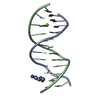 355dS S: Starting model for refinement |
|---|---|
| Similar structure data |


-Links
 PDBj
PDBj


- Assembly
| Deposited unit | 
| ||||||||
|---|---|---|---|---|---|---|---|---|---|
| 1 |
| ||||||||
| Unit cell |
|
-Components
| #1: DNA chain | Mass: 3677.419 Da / Num. of mol.: 2 / Source method: obtained synthetically #2: Chemical | ChemComp-MG / |   Mass: 24.305 Da / Num. of mol.: 1 / Source method: obtained synthetically / Formula: Mg Mass: 24.305 Da / Num. of mol.: 1 / Source method: obtained synthetically / Formula: Mg#3: Water | ChemComp-HOH / |  Mass: 18.015 Da / Num. of mol.: 111 / Source method: isolated from a natural source / Formula: H2O Mass: 18.015 Da / Num. of mol.: 111 / Source method: isolated from a natural source / Formula: H2O |
|---|
-Experimental details
-Experiment
| Experiment | Method: X-RAY DIFFRACTION / Number of used crystals: 1 |
|---|
- Sample preparation
| Crystal | Density Matthews: 2.15 Å3/Da / Density % sol: 42.77 % | ||||||||||||||||||||||||||||||||||||||||||||||||||||||
|---|---|---|---|---|---|---|---|---|---|---|---|---|---|---|---|---|---|---|---|---|---|---|---|---|---|---|---|---|---|---|---|---|---|---|---|---|---|---|---|---|---|---|---|---|---|---|---|---|---|---|---|---|---|---|---|
| Crystal grow | Temperature: 277 K / Method: vapor diffusion, hanging drop / pH: 7 Details: MPD, spermine, magnesium acetate, sodium cacodylate, sodum chloride, pH 7.0, VAPOR DIFFUSION, HANGING DROP, temperature 277K | ||||||||||||||||||||||||||||||||||||||||||||||||||||||
| Components of the solutions |
| ||||||||||||||||||||||||||||||||||||||||||||||||||||||
| Crystal grow | *PLUS Temperature: 4 K | ||||||||||||||||||||||||||||||||||||||||||||||||||||||
| Components of the solutions | *PLUS
|
-Data collection
| Diffraction | Mean temperature: 100 K |
|---|---|
| Diffraction source | Source: SYNCHROTRON / Site: Photon Factory  / Beamline: BL-18B / Wavelength: 1 Å / Beamline: BL-18B / Wavelength: 1 Å |
| Detector | Type: WEISSENBERG / Detector: DIFFRACTOMETER / Date: Dec 12, 2000 |
| Radiation | Protocol: SINGLE WAVELENGTH / Monochromatic (M) / Laue (L): M / Scattering type: x-ray |
| Radiation wavelength | Wavelength: 1 Å / Relative weight: 1 |
| Reflection | Resolution: 1.6→100 Å / Num. all: 62759 / Num. obs: 7855 / % possible obs: 86.1 % / Redundancy: 8 % / Rmerge(I) obs: 0.058 |
| Reflection shell | Resolution: 1.6→1.69 Å / Rmerge(I) obs: 0.132 / Mean I/σ(I) obs: 5.5 / % possible all: 38.4 |
| Reflection | *PLUS Lowest resolution: 100 Å / Redundancy: 5.1 % / Num. measured all: 62759 |
| Reflection shell | *PLUS % possible obs: 38.4 % |
- Processing
| Software |
| ||||||||||||||||
|---|---|---|---|---|---|---|---|---|---|---|---|---|---|---|---|---|---|
| Refinement | Method to determine structure: MOLECULAR REPLACEMENT Starting model: PDB ENTRY 355D Resolution: 1.6→10 Å / Cross valid method: THROUGHOUT / σ(F): 3
| ||||||||||||||||
| Displacement parameters |
| ||||||||||||||||
| Refine analyze | Luzzati coordinate error obs: 0.22 Å / Luzzati d res low obs: 5 Å | ||||||||||||||||
| Refinement step | Cycle: LAST / Resolution: 1.6→10 Å
| ||||||||||||||||
| Refine LS restraints |
| ||||||||||||||||
| LS refinement shell | Resolution: 1.6→1.66 Å / Rfactor Rfree error: 0.124
| ||||||||||||||||
| Software | *PLUS Name: CNS / Version: 1 / Classification: refinement | ||||||||||||||||
| Refinement | *PLUS Highest resolution: 1.6 Å / Lowest resolution: 10 Å / σ(F): 3 / % reflection Rfree: 10 % / Rfactor obs: 0.218 | ||||||||||||||||
| Solvent computation | *PLUS | ||||||||||||||||
| Displacement parameters | *PLUS | ||||||||||||||||
| Refine LS restraints | *PLUS
| ||||||||||||||||
| LS refinement shell | *PLUS Highest resolution: 1.6 Å / Rfactor Rfree: 0.277 / Num. reflection Rfree: 5.2 / Rfactor Rwork: 0.206 |
