 Movie
Movie Controller

[English] 日本語
 Yorodumi
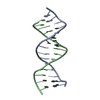
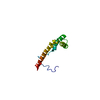
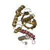
- PDB-1j5n: Solution Structure of the Non-Sequence-Specific HMGB protein NHP6... -
+ Open data

- Basic information
| Entry | Database: PDB / ID: 1j5n | ||||||
|---|---|---|---|---|---|---|---|
| Title | Solution Structure of the Non-Sequence-Specific HMGB protein NHP6A in complex with SRY DNA | ||||||
 Components Components |
| ||||||
 Keywords Keywords | DNA BINDING PROTEIN/DNA / HMG-BOX / HMGB / PROTEIN-DNA COMPLEX / ALPHA HELIX / DOUBLE HELIX / DNA BINDING PROTEIN-DNA COMPLEX | ||||||
| Function / homology |  Function and homology information Function and homology informationmaintenance of transcriptional fidelity during transcription elongation by RNA polymerase III / Regulation of TLR by endogenous ligand / Apoptosis induced DNA fragmentation / Mitochondrial transcription initiation / protein-DNA complex assembly / MutSalpha complex binding / Pyroptosis / RNA polymerase III preinitiation complex assembly / Mitochondrial protein degradation / DNA binding, bending ...maintenance of transcriptional fidelity during transcription elongation by RNA polymerase III / Regulation of TLR by endogenous ligand / Apoptosis induced DNA fragmentation / Mitochondrial transcription initiation / protein-DNA complex assembly / MutSalpha complex binding / Pyroptosis / RNA polymerase III preinitiation complex assembly / Mitochondrial protein degradation / DNA binding, bending / RNA polymerase II preinitiation complex assembly / nucleosome binding / Neutrophil degranulation / protein-DNA complex / chromosome / chromatin organization / nucleic acid binding / chromatin remodeling / DNA repair / nucleus Similarity search - Function | ||||||
| Biological species |  | ||||||
| Method | SOLUTION NMR / simulated annealing | ||||||
 Authors Authors | Masse, J.E. / Wong, B. / Yen, Y.-M. / Allain, F.H.-T. / Johnson, R.C. / Feigon, J. | ||||||
 Citation Citation | Journal: J.Mol.Biol. / Year: 2002 Title: The S. cerevisiae architectural HMGB protein NHP6A complexed with DNA: DNA and protein conformational changes upon binding Authors: Masse, J.E. / Wong, B. / Yen, Y.-M. / Allain, F.H.-T. / Johnson, R.C. / Feigon, J. | ||||||
| History |
|
- Structure visualization
| Structure viewer | Molecule: MolmilJmol/JSmol |
|---|
- Downloads & links
-Download
| PDBx/mmCIF format | 1j5n.cif.gz | 974.6 KB | Display | PDBx/mmCIF format |
|---|---|---|---|---|
| PDB format | pdb1j5n.ent.gz | 804.2 KB | Display | PDB format |
| PDBx/mmJSON format | 1j5n.json.gz | Tree view | PDBx/mmJSON format | |
| Others |  Other downloads Other downloads |
-Validation report
| Arichive directory | https://data.pdbj.org/pub/pdb/validation_reports/j5/1j5nftp://data.pdbj.org/pub/pdb/validation_reports/j5/1j5n | HTTPS FTP |
|---|
-Related structure data


-Links
 PDBj
PDBj


- Assembly
| Deposited unit | 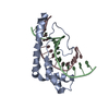
| |||||||||
|---|---|---|---|---|---|---|---|---|---|---|
| 1 |
| |||||||||
| NMR ensembles |
|
-Components
| #1: DNA chain | Mass: 4696.042 Da / Num. of mol.: 1 / Source method: obtained synthetically Details: unlabeled DNA made on a commercial synthesizer, 15N,13C-labeled DNA made enzymatically in vitro with Taq polymerase |
|---|---|
| #2: DNA chain | Mass: 4482.940 Da / Num. of mol.: 1 / Source method: obtained synthetically Details: unlabeled DNA made on a commercial synthesizer, 15N,13C-labeled DNA made enzymatically in vitro with Taq polymerase |
| #3: Protein | Mass: 10824.364 Da / Num. of mol.: 1 Source method: isolated from a genetically manipulated source Source: (gene. exp.) Production host:  |
-Experimental details
-Experiment
| Experiment | Method: SOLUTION NMR | ||||||||||||||||||||||||||||||||||||||||||||||||||||||||||||||||||||||||||||||||
|---|---|---|---|---|---|---|---|---|---|---|---|---|---|---|---|---|---|---|---|---|---|---|---|---|---|---|---|---|---|---|---|---|---|---|---|---|---|---|---|---|---|---|---|---|---|---|---|---|---|---|---|---|---|---|---|---|---|---|---|---|---|---|---|---|---|---|---|---|---|---|---|---|---|---|---|---|---|---|---|---|---|
| NMR experiment |
| ||||||||||||||||||||||||||||||||||||||||||||||||||||||||||||||||||||||||||||||||
| NMR details | Text: This structure calculated using standard 3D heteronuclear filtering and editing techniques with both labeled protein and labeled DNA. |
- Sample preparation
| Details |
| |||||||||||||||
|---|---|---|---|---|---|---|---|---|---|---|---|---|---|---|---|---|
| Sample conditions |
| |||||||||||||||
| Crystal grow | *PLUS Method: other / Details: NMR |
-NMR measurement
| NMR spectrometer | Type: Bruker DRX / Manufacturer: Bruker / Model: DRX / Field strength: 600 MHz |
|---|
- Processing
| NMR software |
| ||||||||||||||||
|---|---|---|---|---|---|---|---|---|---|---|---|---|---|---|---|---|---|
| Refinement | Method: simulated annealing / Software ordinal: 1 Details: The structures are based on a total of 3035 restraints. 102 are from alpha-helical hydrogen bond restraints within the protein (2/h-bond). 76 are hydrogen bond restraints for Watson-Crick ...Details: The structures are based on a total of 3035 restraints. 102 are from alpha-helical hydrogen bond restraints within the protein (2/h-bond). 76 are hydrogen bond restraints for Watson-Crick base pairs in the DNA. The rest are NOE-derived. 2197 of the NOE restraints are protein-protein restraints, 578 are DNA-DNA restraints, and 82 are protein-DNA restraints. | ||||||||||||||||
| NMR representative | Selection criteria: closest to the average | ||||||||||||||||
| NMR ensemble | Conformer selection criteria: structures with acceptable covalent geometry Conformers calculated total number: 100 / Conformers submitted total number: 20 |
 X-PLOR