 Movie
Movie Controller

[English] 日本語
 Yorodumi
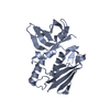
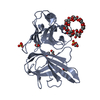
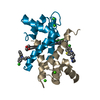
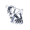
- PDB-1j55: The Crystal Structure of Ca+-bound Human S100P Determined at 2.0A... -
+ Open data

- Basic information
| Entry | Database: PDB / ID: 1j55 | ||||||
|---|---|---|---|---|---|---|---|
| Title | The Crystal Structure of Ca+-bound Human S100P Determined at 2.0A Resolution by X-ray | ||||||
 Components Components | S-100P PROTEIN | ||||||
 Keywords Keywords | METAL BINDING PROTEIN | ||||||
| Function / homology |  Function and homology information Function and homology informationmicrovillus membrane / transition metal ion binding / endothelial cell migration / calcium-dependent protein binding / secretory granule lumen / cadherin binding / calcium ion binding / Neutrophil degranulation / magnesium ion binding / protein homodimerization activity ...microvillus membrane / transition metal ion binding / endothelial cell migration / calcium-dependent protein binding / secretory granule lumen / cadherin binding / calcium ion binding / Neutrophil degranulation / magnesium ion binding / protein homodimerization activity / extracellular exosome / extracellular region / nucleoplasm / nucleus / cytoplasm Similarity search - Function | ||||||
| Biological species |  Homo sapiens (human) Homo sapiens (human) | ||||||
| Method |  X-RAY DIFFRACTION / Resolution: 2 Å X-RAY DIFFRACTION / Resolution: 2 Å | ||||||
 Authors Authors | Zhang, H. / Wang, G. / Ding, Y. / Wang, Z. / Barraclough, R. / Rudland, P.S. / Fernig, D.G. / Rao, Z. | ||||||
 Citation Citation | Journal: J.Mol.Biol. / Year: 2003 Title: The Crystal Structure at 2A Resolution of the Ca2+-binding Protein S100P Authors: Zhang, H. / Wang, G. / Ding, Y. / Wang, Z. / Barraclough, R. / Rudland, P.S. / Fernig, D.G. / Rao, Z. | ||||||
| History |
|
- Structure visualization
| Structure viewer | Molecule: MolmilJmol/JSmol |
|---|
- Downloads & links
-Download
| PDBx/mmCIF format | 1j55.cif.gz | 29.8 KB | Display | PDBx/mmCIF format |
|---|---|---|---|---|
| PDB format | pdb1j55.ent.gz | 19.2 KB | Display | PDB format |
| PDBx/mmJSON format | 1j55.json.gz | Tree view | PDBx/mmJSON format | |
| Others |  Other downloads Other downloads |
-Validation report
| Arichive directory | https://data.pdbj.org/pub/pdb/validation_reports/j5/1j55ftp://data.pdbj.org/pub/pdb/validation_reports/j5/1j55 | HTTPS FTP |
|---|
-Related structure data
| Similar structure data |
|---|


-Links
 PDBj
PDBj- Assembly
| Deposited unit | 
| ||||||||
|---|---|---|---|---|---|---|---|---|---|
| 1 | 
| ||||||||
| Unit cell |
|
-Components
| #1: Protein | Mass: 10411.884 Da / Num. of mol.: 1 Source method: isolated from a genetically manipulated source Source: (gene. exp.) Homo sapiens (human) / Plasmid: PET11a / Production host:  | ||
|---|---|---|---|
| #2: Chemical |   Mass: 40.078 Da / Num. of mol.: 2 / Source method: obtained synthetically / Formula: Ca Mass: 40.078 Da / Num. of mol.: 2 / Source method: obtained synthetically / Formula: Ca#3: Water | ChemComp-HOH / |  Mass: 18.015 Da / Num. of mol.: 46 / Source method: isolated from a natural source / Formula: H2O Mass: 18.015 Da / Num. of mol.: 46 / Source method: isolated from a natural source / Formula: H2O |
-Experimental details
-Experiment
| Experiment | Method: X-RAY DIFFRACTION / Number of used crystals: 1 |
|---|
- Sample preparation
| Crystal | Density Matthews: 2.11 Å3/Da / Density % sol: 41.83 % | |||||||||||||||||||||||||||||||||||
|---|---|---|---|---|---|---|---|---|---|---|---|---|---|---|---|---|---|---|---|---|---|---|---|---|---|---|---|---|---|---|---|---|---|---|---|---|
| Crystal grow | Temperature: 291 K / Method: evaporation / pH: 6.5 Details: PEG4000, sodium citrate, calcium chloride, pH 6.5, EVAPORATION, temperature 291K | |||||||||||||||||||||||||||||||||||
| Crystal grow | *PLUS Method: vapor diffusion, hanging drop | |||||||||||||||||||||||||||||||||||
| Components of the solutions | *PLUS
|
-Data collection
| Diffraction | Mean temperature: 110 K |
|---|---|
| Diffraction source | Source: ROTATING ANODE / Type: RIGAKU RU200 / Wavelength: 1.5418 |
| Detector | Type: MAR scanner 345 mm plate / Detector: IMAGE PLATE / Date: Oct 11, 2001 |
| Radiation | Protocol: SINGLE WAVELENGTH / Monochromatic (M) / Laue (L): M / Scattering type: x-ray |
| Radiation wavelength | Wavelength: 1.5418 Å / Relative weight: 1 |
| Reflection | Resolution: 2→40 Å / Num. all: 57925 / Num. obs: 57916 / % possible obs: 96.6 % / Observed criterion σ(F): 0 / Observed criterion σ(I): -3 / Redundancy: 8.9 % / Biso Wilson estimate: 11.5 Å2 / Rmerge(I) obs: 0.104 / Net I/σ(I): 14.9 |
| Reflection shell | Resolution: 2→2.07 Å / Redundancy: 8.1 % / Rmerge(I) obs: 0.403 / Num. unique all: 615 / % possible all: 96.6 |
| Reflection | *PLUS Lowest resolution: 50 Å / Num. obs: 6442 / % possible obs: 100 % |
| Reflection shell | *PLUS Highest resolution: 2 Å / % possible obs: 100 % / Num. unique obs: 615 / Mean I/σ(I) obs: 58.2 |
- Processing
| Software |
| |||||||||||||||||||||||||
|---|---|---|---|---|---|---|---|---|---|---|---|---|---|---|---|---|---|---|---|---|---|---|---|---|---|---|
| Refinement | Resolution: 2→40 Å / Cross valid method: THROUGHOUT / σ(F): 0 / σ(I): 0 / Stereochemistry target values: Engh & Huber / Details: CNS
| |||||||||||||||||||||||||
| Refinement step | Cycle: LAST / Resolution: 2→40 Å
| |||||||||||||||||||||||||
| Refine LS restraints |
| |||||||||||||||||||||||||
| Refinement | *PLUS Lowest resolution: 40 Å | |||||||||||||||||||||||||
| Solvent computation | *PLUS | |||||||||||||||||||||||||
| Displacement parameters | *PLUS | |||||||||||||||||||||||||
| LS refinement shell | *PLUS Highest resolution: 2 Å / Lowest resolution: 2.07 Å / Rfactor Rfree: 0.281 / Rfactor Rwork: 0.225 |
