 Movie
Movie Controller


+ Open data

- Basic information
| Entry | Database: PDB / ID: 1j37 | ||||||
|---|---|---|---|---|---|---|---|
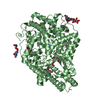
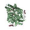
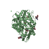
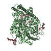
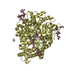
| Title | Crystal Structure of Drosophila AnCE | ||||||
 Components Components | angiotensin converting enzyme | ||||||
 Keywords Keywords | HYDROLASE / Angiotensin | ||||||
| Function / homology |  Function and homology information Function and homology informationMetabolism of Angiotensinogen to Angiotensins / metamorphosis / mating / response to symbiotic bacterium / peptidyl-dipeptidase A / peptide hormone processing / peptidyl-dipeptidase activity / carboxypeptidase activity / metallopeptidase activity / proteolysis ...Metabolism of Angiotensinogen to Angiotensins / metamorphosis / mating / response to symbiotic bacterium / peptidyl-dipeptidase A / peptide hormone processing / peptidyl-dipeptidase activity / carboxypeptidase activity / metallopeptidase activity / proteolysis / : / metal ion binding / plasma membrane Similarity search - Function | ||||||
| Biological species |  | ||||||
| Method |  X-RAY DIFFRACTION / SYNCHROTRON / MIR / Resolution: 2.4 Å X-RAY DIFFRACTION / SYNCHROTRON / MIR / Resolution: 2.4 Å | ||||||
 Authors Authors | Kim, H.M. / Shin, D.R. / Yoo, O.J. / Lee, H. / Lee, J.-O. | ||||||
 Citation Citation | Journal: Febs Lett. / Year: 2003 Title: Crystal structure of Drosophila angiotensin I-converting enzyme bound to captopril and lisinopril Authors: Kim, H.M. / Shin, D.R. / Yoo, O.J. / Lee, H. / Lee, J.-O. | ||||||
| History |
|
- Structure visualization
| Structure viewer | Molecule: MolmilJmol/JSmol |
|---|
- Downloads & links
-Download
| PDBx/mmCIF format | 1j37.cif.gz | 249.4 KB | Display | PDBx/mmCIF format |
|---|---|---|---|---|
| PDB format | pdb1j37.ent.gz | 199.6 KB | Display | PDB format |
| PDBx/mmJSON format | 1j37.json.gz | Tree view | PDBx/mmJSON format | |
| Others |  Other downloads Other downloads |
-Validation report
| Arichive directory | https://data.pdbj.org/pub/pdb/validation_reports/j3/1j37ftp://data.pdbj.org/pub/pdb/validation_reports/j3/1j37 | HTTPS FTP |
|---|
-Related structure data


-Links
 PDBj
PDBj


- Assembly
| Deposited unit | 
| ||||||||
|---|---|---|---|---|---|---|---|---|---|
| 1 | 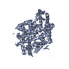
| ||||||||
| 2 | 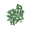
| ||||||||
| Unit cell |
|
-Components
| #1: Protein | Mass: 70440.156 Da / Num. of mol.: 2 Source method: isolated from a genetically manipulated source Source: (gene. exp.)  Spodoptera frugiperda (fall armyworm) / References: UniProt: Q10714, peptidyl-dipeptidase A Spodoptera frugiperda (fall armyworm) / References: UniProt: Q10714, peptidyl-dipeptidase A#2: Chemical |   Mass: 65.409 Da / Num. of mol.: 2 / Source method: obtained synthetically / Formula: Zn Mass: 65.409 Da / Num. of mol.: 2 / Source method: obtained synthetically / Formula: Zn#3: Chemical |   Mass: 217.285 Da / Num. of mol.: 2 / Source method: obtained synthetically / Formula: C9H15NO3S / Comment: inhibitor, medication*YM Mass: 217.285 Da / Num. of mol.: 2 / Source method: obtained synthetically / Formula: C9H15NO3S / Comment: inhibitor, medication*YMHas protein modification | Y | |
|---|
-Experimental details
-Experiment
| Experiment | Method: X-RAY DIFFRACTION / Number of used crystals: 1 |
|---|
- Sample preparation
| Crystal | Density Matthews: 3.82 Å3/Da / Density % sol: 67.77 % | ||||||||||||||||||||||||||||||||||||||||||
|---|---|---|---|---|---|---|---|---|---|---|---|---|---|---|---|---|---|---|---|---|---|---|---|---|---|---|---|---|---|---|---|---|---|---|---|---|---|---|---|---|---|---|---|
| Crystal grow | Temperature: 293 K / Method: vapor diffusion, hanging drop / pH: 7.5 Details: Sodium Formate 7.2M, pH 7.5, VAPOR DIFFUSION, HANGING DROP, temperature 293K | ||||||||||||||||||||||||||||||||||||||||||
| Crystal grow | *PLUS Temperature: 22 ℃ / Method: vapor diffusion, hanging drop | ||||||||||||||||||||||||||||||||||||||||||
| Components of the solutions | *PLUS
|
-Data collection
| Diffraction | Mean temperature: 123 K |
|---|---|
| Diffraction source | Source: SYNCHROTRON / Site: CHESS  / Beamline: A1 / Wavelength: 0.928 Å / Beamline: A1 / Wavelength: 0.928 Å |
| Detector | Type: ADSC QUANTUM 4 / Detector: CCD / Date: Dec 22, 2000 |
| Radiation | Monochromator: Mirror / Protocol: SINGLE WAVELENGTH / Monochromatic (M) / Laue (L): M / Scattering type: x-ray |
| Radiation wavelength | Wavelength: 0.928 Å / Relative weight: 1 |
| Reflection | Resolution: 2.4→20 Å / Num. all: 91258 / Num. obs: 82133 / % possible obs: 90.8 % / Observed criterion σ(F): 0 / Observed criterion σ(I): 0 |
| Reflection shell | Resolution: 2.4→2.5 Å / % possible all: 76.4 |
| Reflection | *PLUS Num. measured all: 590560 / Rmerge(I) obs: 0.059 |
| Reflection shell | *PLUS % possible obs: 76.4 % / Rmerge(I) obs: 0.375 / Mean I/σ(I) obs: 3 |
- Processing
| Software |
| ||||||||||||||||||||
|---|---|---|---|---|---|---|---|---|---|---|---|---|---|---|---|---|---|---|---|---|---|
| Refinement | Method to determine structure: MIR / Resolution: 2.4→20 Å / σ(F): 0 / σ(I): 0 / Stereochemistry target values: Engh & Huber
| ||||||||||||||||||||
| Refinement step | Cycle: LAST / Resolution: 2.4→20 Å
| ||||||||||||||||||||
| Refinement | *PLUS Lowest resolution: 20 Å | ||||||||||||||||||||
| Solvent computation | *PLUS | ||||||||||||||||||||
| Displacement parameters | *PLUS | ||||||||||||||||||||
| Refine LS restraints | *PLUS
|
