 Movie
Movie Controller

[English] 日本語
 Yorodumi
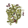
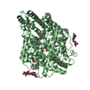
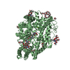
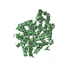
- PDB-5ama: Crystal structure of the Angiotensin-1 converting enzyme N-domain... -
+ Open data

- Basic information
| Entry | Database: PDB / ID: 5ama | |||||||||
|---|---|---|---|---|---|---|---|---|---|---|
| Title | Crystal structure of the Angiotensin-1 converting enzyme N-domain in complex with amyloid-beta 1-16 | |||||||||
 Components Components | ANGIOTENSIN-CONVERTING ENZYME | |||||||||
 Keywords Keywords | HYDROLASE / METALLOPROTEASE / AMYLOID- BETA | |||||||||
| Function / homology |  Function and homology information Function and homology informationmononuclear cell proliferation / cell proliferation in bone marrow / bradykinin receptor binding / exopeptidase activity / regulation of angiotensin metabolic process / substance P catabolic process / tripeptidyl-peptidase activity / peptidyl-dipeptidase A / regulation of renal output by angiotensin / negative regulation of gap junction assembly ...mononuclear cell proliferation / cell proliferation in bone marrow / bradykinin receptor binding / exopeptidase activity / regulation of angiotensin metabolic process / substance P catabolic process / tripeptidyl-peptidase activity / peptidyl-dipeptidase A / regulation of renal output by angiotensin / negative regulation of gap junction assembly / hormone catabolic process / bradykinin catabolic process / hormone metabolic process / metallodipeptidase activity / regulation of smooth muscle cell migration / regulation of hematopoietic stem cell proliferation / neutrophil mediated immunity / mitogen-activated protein kinase binding / mitogen-activated protein kinase kinase binding / chloride ion binding / arachidonate secretion / post-transcriptional regulation of gene expression / peptide catabolic process / heart contraction / positive regulation of systemic arterial blood pressure / regulation of heart rate by cardiac conduction / amyloid-beta metabolic process / hematopoietic stem cell differentiation / regulation of systemic arterial blood pressure by renin-angiotensin / blood vessel remodeling / peptidyl-dipeptidase activity / antigen processing and presentation of peptide antigen via MHC class I / regulation of vasoconstriction / Metabolism of Angiotensinogen to Angiotensins / angiotensin maturation / metallocarboxypeptidase activity / blood vessel diameter maintenance / angiotensin-activated signaling pathway / kidney development / metalloendopeptidase activity / male gonad development / regulation of synaptic plasticity / regulation of blood pressure / metallopeptidase activity / peptidase activity / actin binding / endopeptidase activity / spermatogenesis / calmodulin binding / lysosome / endosome / external side of plasma membrane / negative regulation of gene expression / proteolysis / : / extracellular exosome / extracellular region / zinc ion binding / plasma membrane Similarity search - Function | |||||||||
| Biological species |  HOMO SAPIENS (human) HOMO SAPIENS (human) | |||||||||
| Method |  X-RAY DIFFRACTION / SYNCHROTRON / MOLECULAR REPLACEMENT / Resolution: 1.8 Å X-RAY DIFFRACTION / SYNCHROTRON / MOLECULAR REPLACEMENT / Resolution: 1.8 Å | |||||||||
 Authors Authors | Masuyer, G. / Larmuth, K.M. / Douglas, R.G. / Sturrock, E.D. / Acharya, K.R. | |||||||||
 Citation Citation | Journal: FEBS J. / Year: 2016 Title: The Kinetic and Structural Characterisation of Amyloid-Beta Metabolism by Human Angiotensin-1- Converting Enzyme (Ace) Authors: Larmuth, K.M. / Masuyer, G. / Douglas, R.G. / Sturrock, E.D. / Acharya, K.R. | |||||||||
| History |
|
- Structure visualization
| Structure viewer | Molecule: MolmilJmol/JSmol |
|---|
- Downloads & links
-Download
| PDBx/mmCIF format | 5ama.cif.gz | 1009.9 KB | Display | PDBx/mmCIF format |
|---|---|---|---|---|
| PDB format | pdb5ama.ent.gz | 844.2 KB | Display | PDB format |
| PDBx/mmJSON format | 5ama.json.gz | Tree view | PDBx/mmJSON format | |
| Others |  Other downloads Other downloads |
-Validation report
| Arichive directory | https://data.pdbj.org/pub/pdb/validation_reports/am/5amaftp://data.pdbj.org/pub/pdb/validation_reports/am/5ama | HTTPS FTP |
|---|
-Related structure data
| Related structure data |  5am8C 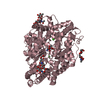 5am9C 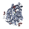 5ambC  5amcC  3nxqS C: citing same article ( S: Starting model for refinement |
|---|---|
| Similar structure data |


-Links
 PDBj
PDBj


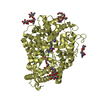
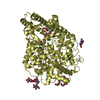
- Assembly
| Deposited unit | 
| ||||||||
|---|---|---|---|---|---|---|---|---|---|
| 1 | 
| ||||||||
| 2 | 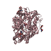
| ||||||||
| 3 | 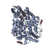
| ||||||||
| 4 | 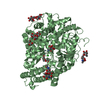
| ||||||||
| Unit cell |
|
-Components
-Protein , 1 types, 4 molecules ABCD
| #1: Protein | Mass: 72606.508 Da / Num. of mol.: 4 / Fragment: N DOMAIN, UNP RESIDUES 30-658 / Mutation: YES Source method: isolated from a genetically manipulated source Details: MINIMALLY GLYCOSYLATED MUTANT / Source: (gene. exp.) HOMO SAPIENS (human) / Cell line (production host): CHO K1 / Production host:   CRICETULUS GRISEUS (Chinese hamster) / References: UniProt: P12821, peptidyl-dipeptidase A CRICETULUS GRISEUS (Chinese hamster) / References: UniProt: P12821, peptidyl-dipeptidase A |
|---|
-Sugars , 6 types, 12 molecules 
| #2: Polysaccharide | 2-acetamido-2-deoxy-beta-D-glucopyranose-(1-4)-2-acetamido-2-deoxy-beta-D-glucopyranose Source method: isolated from a genetically manipulated source #3: Polysaccharide | Source method: isolated from a genetically manipulated source #4: Polysaccharide | beta-D-mannopyranose-(1-4)-2-acetamido-2-deoxy-beta-D-glucopyranose-(1-4)-2-acetamido-2-deoxy-beta- ...beta-D-mannopyranose-(1-4)-2-acetamido-2-deoxy-beta-D-glucopyranose-(1-4)-2-acetamido-2-deoxy-beta-D-glucopyranose | Source method: isolated from a genetically manipulated source #5: Polysaccharide | beta-D-mannopyranose-(1-4)-2-acetamido-2-deoxy-beta-D-glucopyranose-(1-4)-[beta-L-fucopyranose-(1-6) ...beta-D-mannopyranose-(1-4)-2-acetamido-2-deoxy-beta-D-glucopyranose-(1-4)-[beta-L-fucopyranose-(1-6)]2-acetamido-2-deoxy-beta-D-glucopyranose | Source method: isolated from a genetically manipulated source #6: Polysaccharide | alpha-D-mannopyranose-(1-4)-2-acetamido-2-deoxy-beta-D-glucopyranose-(1-4)-[alpha-L-fucopyranose-(1- ...alpha-D-mannopyranose-(1-4)-2-acetamido-2-deoxy-beta-D-glucopyranose-(1-4)-[alpha-L-fucopyranose-(1-6)]2-acetamido-2-deoxy-beta-D-glucopyranose | Source method: isolated from a genetically manipulated source #11: Sugar |  Type: D-saccharide, beta linking / Mass: 221.208 Da / Num. of mol.: 2 Type: D-saccharide, beta linking / Mass: 221.208 Da / Num. of mol.: 2Source method: isolated from a genetically manipulated source Formula: C8H15NO6 |
|---|
-Non-polymers , 7 types, 1923 molecules 


| #7: Chemical | ChemComp-ASP /  Type: L-peptide linking / Mass: 133.103 Da / Num. of mol.: 4 / Source method: obtained synthetically / Formula: C4H7NO4 Type: L-peptide linking / Mass: 133.103 Da / Num. of mol.: 4 / Source method: obtained synthetically / Formula: C4H7NO4#8: Chemical | ChemComp-SER /  Type: L-peptide linking / Mass: 105.093 Da / Num. of mol.: 4 / Source method: obtained synthetically / Formula: C3H7NO3 Type: L-peptide linking / Mass: 105.093 Da / Num. of mol.: 4 / Source method: obtained synthetically / Formula: C3H7NO3#9: Chemical | ChemComp-ZN /  Mass: 65.409 Da / Num. of mol.: 4 / Source method: obtained synthetically / Formula: Zn Mass: 65.409 Da / Num. of mol.: 4 / Source method: obtained synthetically / Formula: Zn#10: Chemical | ChemComp-CL /  Mass: 35.453 Da / Num. of mol.: 4 / Source method: obtained synthetically / Formula: Cl Mass: 35.453 Da / Num. of mol.: 4 / Source method: obtained synthetically / Formula: Cl#12: Chemical | ChemComp-PEG /  Mass: 106.120 Da / Num. of mol.: 11 / Source method: obtained synthetically / Formula: C4H10O3 Mass: 106.120 Da / Num. of mol.: 11 / Source method: obtained synthetically / Formula: C4H10O3#13: Chemical | ChemComp-P6G /  Mass: 282.331 Da / Num. of mol.: 6 / Source method: obtained synthetically / Formula: C12H26O7 / Comment: precipitant*YM Mass: 282.331 Da / Num. of mol.: 6 / Source method: obtained synthetically / Formula: C12H26O7 / Comment: precipitant*YM#14: Water | ChemComp-HOH / | Mass: 18.015 Da / Num. of mol.: 1890 / Source method: isolated from a natural source / Formula: H2O |
|---|
-Details
| Has protein modification | Y |
|---|
-Experimental details
-Experiment
| Experiment | Method: X-RAY DIFFRACTION / Number of used crystals: 1 |
|---|
- Sample preparation
| Crystal | Density Matthews: 2.95 Å3/Da / Density % sol: 58 % / Description: NONE |
|---|---|
| Crystal grow | pH: 8.5 Details: 0.06 M DIVALENT CATIONS, 0.1 M TRIS/BICINE PH 8.5, 30 % PEG550MME/PEG20000 |
-Data collection
| Diffraction | Mean temperature: 100 K |
|---|---|
| Diffraction source | Source: SYNCHROTRON / Site: Diamond  / Beamline: I03 / Wavelength: 0.9763 / Beamline: I03 / Wavelength: 0.9763 |
| Detector | Type: DECTRIS PILATUS 6M / Detector: PIXEL / Date: Oct 13, 2014 |
| Radiation | Protocol: SINGLE WAVELENGTH / Monochromatic (M) / Laue (L): M / Scattering type: x-ray |
| Radiation wavelength | Wavelength: 0.9763 Å / Relative weight: 1 |
| Reflection | Resolution: 1.8→33.42 Å / Num. obs: 281258 / % possible obs: 93.6 % / Observed criterion σ(I): 0 / Redundancy: 1.6 % / Rmerge(I) obs: 0.06 / Net I/σ(I): 6.8 |
| Reflection shell | Resolution: 1.8→1.9 Å / Redundancy: 1.5 % / Rmerge(I) obs: 0.46 / Mean I/σ(I) obs: 1.4 / % possible all: 82.1 |
- Processing
| Software |
| ||||||||||||||||||||||||||||||||||||||||||||||||||||||||||||||||||||||||||||||||||||||||||||||||||||||||||||||||||||||||||||||||||||||||||||||||||||||||||||||||||||||||||||||||||||||
|---|---|---|---|---|---|---|---|---|---|---|---|---|---|---|---|---|---|---|---|---|---|---|---|---|---|---|---|---|---|---|---|---|---|---|---|---|---|---|---|---|---|---|---|---|---|---|---|---|---|---|---|---|---|---|---|---|---|---|---|---|---|---|---|---|---|---|---|---|---|---|---|---|---|---|---|---|---|---|---|---|---|---|---|---|---|---|---|---|---|---|---|---|---|---|---|---|---|---|---|---|---|---|---|---|---|---|---|---|---|---|---|---|---|---|---|---|---|---|---|---|---|---|---|---|---|---|---|---|---|---|---|---|---|---|---|---|---|---|---|---|---|---|---|---|---|---|---|---|---|---|---|---|---|---|---|---|---|---|---|---|---|---|---|---|---|---|---|---|---|---|---|---|---|---|---|---|---|---|---|---|---|---|---|
| Refinement | Method to determine structure: MOLECULAR REPLACEMENT Starting model: PDB ENTRY 3NXQ Resolution: 1.8→113.48 Å / Cor.coef. Fo:Fc: 0.96 / Cor.coef. Fo:Fc free: 0.945 / SU B: 7.095 / SU ML: 0.092 / Cross valid method: THROUGHOUT / ESU R: 0.124 / ESU R Free: 0.118 / Stereochemistry target values: MAXIMUM LIKELIHOOD Details: HYDROGENS HAVE BEEN ADDED IN THE RIDING POSITIONS. ONLY DI-PEPTIDE ASP-SER (7-8) VISIBLE FOR AMYLOID-BETA ( PRODUCT OF CLEAVAGE REACTION BY ACE)
| ||||||||||||||||||||||||||||||||||||||||||||||||||||||||||||||||||||||||||||||||||||||||||||||||||||||||||||||||||||||||||||||||||||||||||||||||||||||||||||||||||||||||||||||||||||||
| Solvent computation | Ion probe radii: 0.8 Å / Shrinkage radii: 0.8 Å / VDW probe radii: 1.2 Å / Solvent model: MASK | ||||||||||||||||||||||||||||||||||||||||||||||||||||||||||||||||||||||||||||||||||||||||||||||||||||||||||||||||||||||||||||||||||||||||||||||||||||||||||||||||||||||||||||||||||||||
| Displacement parameters | Biso mean: 23.805 Å2
| ||||||||||||||||||||||||||||||||||||||||||||||||||||||||||||||||||||||||||||||||||||||||||||||||||||||||||||||||||||||||||||||||||||||||||||||||||||||||||||||||||||||||||||||||||||||
| Refinement step | Cycle: LAST / Resolution: 1.8→113.48 Å
| ||||||||||||||||||||||||||||||||||||||||||||||||||||||||||||||||||||||||||||||||||||||||||||||||||||||||||||||||||||||||||||||||||||||||||||||||||||||||||||||||||||||||||||||||||||||
| Refine LS restraints |
|