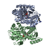
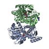
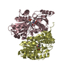
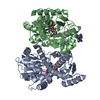
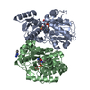
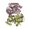
 Movie
Movie Controller


+ Open data

- Basic information
| Entry | Database: PDB / ID: 1j0e | |||||||||
|---|---|---|---|---|---|---|---|---|---|---|
| Title | ACC deaminase mutant reacton intermediate | |||||||||
 Components Components | 1-aminocyclopropane-1-carboxylate deaminase | |||||||||
 Keywords Keywords | LYASE / PLP dependent B group | |||||||||
| Function / homology |  Function and homology information Function and homology informationamine catabolic process / 1-aminocyclopropane-1-carboxylate deaminase / 1-aminocyclopropane-1-carboxylate deaminase activity / D-cysteine desulfhydrase activity / pyridoxal phosphate binding Similarity search - Function | |||||||||
| Biological species |  Williopsis saturnus (fungus) Williopsis saturnus (fungus) | |||||||||
| Method |  X-RAY DIFFRACTION / SYNCHROTRON / FOURIER SYNTHESIS / Resolution: 2.45 Å X-RAY DIFFRACTION / SYNCHROTRON / FOURIER SYNTHESIS / Resolution: 2.45 Å | |||||||||
 Authors Authors | Ose, T. / Fujino, A. / Yao, M. / Honma, M. / Tanaka, I. | |||||||||
 Citation Citation | Journal: J.BIOL.CHEM. / Year: 2003 Title: Reaction intermediate structures of 1-aminocyclopropane-1-carboxylate deaminase: insight into PLP-dependent cyclopropane ring-opening reaction Authors: Ose, T. / Fujino, A. / Yao, M. / Watanabe, N. / Honma, M. / Tanaka, I. #1: Journal: J.Biol.Chem. / Year: 2000Title: Crystal structure of 1-aminocyclopropane-1-carboxylate deaminase from Hansenula saturnus Authors: Yao, M. / Ose, T. / Sugimoto, H. / Horiuchi, A. / Nakagawa, A. / Wakatsuki, S. / Yokoi, D. / Murakami, T. / Honma, M. / Tanaka, I. | |||||||||
| History |
|
- Structure visualization
| Structure viewer | Molecule: MolmilJmol/JSmol |
|---|
- Downloads & links
-Download
| PDBx/mmCIF format | 1j0e.cif.gz | 290.8 KB | Display | PDBx/mmCIF format |
|---|---|---|---|---|
| PDB format | pdb1j0e.ent.gz | 234.4 KB | Display | PDB format |
| PDBx/mmJSON format | 1j0e.json.gz | Tree view | PDBx/mmJSON format | |
| Others |  Other downloads Other downloads |
-Validation report
| Summary document | 1j0e_validation.pdf.gz | 414.7 KB | Display | wwPDB validaton report |
|---|---|---|---|---|
| Full document | 1j0e_full_validation.pdf.gz | 470.2 KB | Display | |
| Data in XML | 1j0e_validation.xml.gz | 37.3 KB | Display | |
| Data in CIF | 1j0e_validation.cif.gz | 56.9 KB | Display | |
| Arichive directory | https://data.pdbj.org/pub/pdb/validation_reports/j0/1j0eftp://data.pdbj.org/pub/pdb/validation_reports/j0/1j0e | HTTPS FTP |
-Related structure data
| Related structure data | 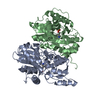 1j0cC 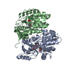 1j0dC 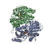 1f2dS S: Starting model for refinement C: citing same article ( |
|---|---|
| Similar structure data |


-Links
 PDBj
PDBj
- Assembly
| Deposited unit | 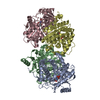
| ||||||||
|---|---|---|---|---|---|---|---|---|---|
| 1 | 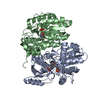
| ||||||||
| 2 | 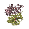
| ||||||||
| Unit cell |
|
-Components
| #1: Protein | Mass: 37299.242 Da / Num. of mol.: 4 / Mutation: S1A, Y295F Source method: isolated from a genetically manipulated source Source: (gene. exp.) Williopsis saturnus (fungus) / Plasmid: pET11d / Species (production host): Escherichia coli / Production host:  References: UniProt: Q7M523, 1-aminocyclopropane-1-carboxylate deaminase #2: Chemical | ChemComp-1AC /   Type: peptide linking / Mass: 101.104 Da / Num. of mol.: 4 / Source method: obtained synthetically / Formula: C4H7NO2 Type: peptide linking / Mass: 101.104 Da / Num. of mol.: 4 / Source method: obtained synthetically / Formula: C4H7NO2#3: Water | ChemComp-HOH / |  Mass: 18.015 Da / Num. of mol.: 939 / Source method: isolated from a natural source / Formula: H2O Mass: 18.015 Da / Num. of mol.: 939 / Source method: isolated from a natural source / Formula: H2OHas protein modification | Y | |
|---|
-Experimental details
-Experiment
| Experiment | Method: X-RAY DIFFRACTION / Number of used crystals: 1 |
|---|
- Sample preparation
| Crystal | Density Matthews: 2.78 Å3/Da / Density % sol: 55.7 % | ||||||||||||||||||||||||||||||||||||||||||||||||
|---|---|---|---|---|---|---|---|---|---|---|---|---|---|---|---|---|---|---|---|---|---|---|---|---|---|---|---|---|---|---|---|---|---|---|---|---|---|---|---|---|---|---|---|---|---|---|---|---|---|
| Crystal grow | Temperature: 293 K / Method: vapor diffusion, hanging drop / pH: 7 Details: ammmonium sulfate, pottasium phosphate, pH 7.0, VAPOR DIFFUSION, HANGING DROP, temperature 293K | ||||||||||||||||||||||||||||||||||||||||||||||||
| Crystal grow | *PLUS pH: 6.5 | ||||||||||||||||||||||||||||||||||||||||||||||||
| Components of the solutions | *PLUS
|
-Data collection
| Diffraction | Mean temperature: 100 K |
|---|---|
| Diffraction source | Source: SYNCHROTRON / Site: SPring-8  / Beamline: BL41XU / Wavelength: 1 Å / Beamline: BL41XU / Wavelength: 1 Å |
| Detector | Type: MARRESEARCH / Detector: CCD / Date: Feb 1, 2002 / Details: mirrors |
| Radiation | Monochromator: GRAPHITE / Protocol: SINGLE WAVELENGTH / Monochromatic (M) / Laue (L): M / Scattering type: x-ray |
| Radiation wavelength | Wavelength: 1 Å / Relative weight: 1 |
| Reflection | Resolution: 2.45→38.3 Å / Num. all: 63065 / Num. obs: 60876 / % possible obs: 99.4 % / Observed criterion σ(F): 2 / Observed criterion σ(I): 2 / Redundancy: 5 % / Biso Wilson estimate: 41.575 Å2 / Rmerge(I) obs: 0.076 / Rsym value: 0.068 / Net I/σ(I): 9.8 |
| Reflection shell | Resolution: 2.45→2.58 Å / Redundancy: 4.7 % / Rmerge(I) obs: 0.368 / Mean I/σ(I) obs: 2.2 / Num. unique all: 8689 / Rsym value: 0.324 / % possible all: 98.6 |
| Reflection | *PLUS Lowest resolution: 38 Å / Num. measured all: 302793 |
| Reflection shell | *PLUS % possible obs: 98.6 % / Num. unique obs: 8689 / Num. measured obs: 41102 |
- Processing
| Software |
| |||||||||||||||||||||||||
|---|---|---|---|---|---|---|---|---|---|---|---|---|---|---|---|---|---|---|---|---|---|---|---|---|---|---|
| Refinement | Method to determine structure: FOURIER SYNTHESIS Starting model: PDB ENTRY 1F2D Resolution: 2.45→10 Å / Isotropic thermal model: Isotropic / Cross valid method: THROUGHOUT / σ(F): 0 / σ(I): 0 / Stereochemistry target values: Engh & Huber
| |||||||||||||||||||||||||
| Displacement parameters | Biso mean: 45.1457 Å2
| |||||||||||||||||||||||||
| Refine analyze |
| |||||||||||||||||||||||||
| Refinement step | Cycle: LAST / Resolution: 2.45→10 Å
| |||||||||||||||||||||||||
| Refine LS restraints |
| |||||||||||||||||||||||||
| LS refinement shell | Resolution: 2.45→2.54 Å
| |||||||||||||||||||||||||
| Refinement | *PLUS Highest resolution: 2.45 Å / Lowest resolution: 38 Å | |||||||||||||||||||||||||
| Solvent computation | *PLUS | |||||||||||||||||||||||||
| Displacement parameters | *PLUS | |||||||||||||||||||||||||
| Refine LS restraints | *PLUS
|
