 Movie
Movie Controller

[English] 日本語
 Yorodumi
- PDB-1ixb: CRYSTAL STRUCTURE OF THE E. COLI MANGANESE(II) SUPEROXIDE DISMUTA... -
+ Open data

- Basic information
| Entry | Database: PDB / ID: 1ixb | ||||||
|---|---|---|---|---|---|---|---|
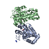
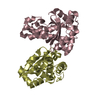
| Title | CRYSTAL STRUCTURE OF THE E. COLI MANGANESE(II) SUPEROXIDE DISMUTASE MUTANT Y174F AT 0.90 ANGSTROMS RESOLUTION. | ||||||
 Components Components | SUPEROXIDE DISMUTASE | ||||||
 Keywords Keywords | OXIDOREDUCTASE / MANGANESE(II) SUPEROXIDE DISMUTASE / Y174F MUTANT / HYDROGEN BOND REACTIVITY / ULTRAHIGH RESOLUTION | ||||||
| Function / homology |  Function and homology information Function and homology informationcellular response to selenium ion / response to acidic pH / superoxide metabolic process / superoxide dismutase / superoxide dismutase activity / antioxidant activity / removal of superoxide radicals / manganese ion binding / response to heat / response to oxidative stress ...cellular response to selenium ion / response to acidic pH / superoxide metabolic process / superoxide dismutase / superoxide dismutase activity / antioxidant activity / removal of superoxide radicals / manganese ion binding / response to heat / response to oxidative stress / protein homodimerization activity / DNA binding / metal ion binding / cytoplasm / cytosol Similarity search - Function | ||||||
| Biological species |  | ||||||
| Method |  X-RAY DIFFRACTION / SYNCHROTRON / MOLECULAR REPLACEMENT / Resolution: 0.9 Å X-RAY DIFFRACTION / SYNCHROTRON / MOLECULAR REPLACEMENT / Resolution: 0.9 Å | ||||||
 Authors Authors | Anderson, B.F. / Edwards, R.A. / Whittaker, M.M. / Whittaker, J.W. / Baker, E.N. / Jameson, G.B. | ||||||
 Citation Citation | Journal: To be Published Title: Structures at 0.90 A resolution of the oxidised and reduced forms of the Y174F mutant of the manganese superoxide dismutase from Escherichia coli Authors: Anderson, B.F. / Edwards, R.A. / Whittaker, M.M. / Whittaker, J.W. / Baker, E.N. / Jameson, G.B. #1: Journal: Biochemistry / Year: 2001Title: Removing a Hydrogen Bond in the Dimer Interface of Escherichia Coli Manganese Superoxide Dismutase Alters Structure and Reactivity Authors: Edwards, R.A. / Whittaker, M.M. / Whittaker, J.W. / Baker, E.N. / Jameson, G.B. #2: Journal: Biochemistry / Year: 2001Title: Outer Sphere Mutations Perturb Metal Reactivity in Manganese Superoxide Dismutase Authors: Edwards, R.A. / Whittaker, M.M. / Whittaker, J.W. / Baker, E.N. / Jameson, G.B. | ||||||
| History |
|
- Structure visualization
| Structure viewer | Molecule: MolmilJmol/JSmol |
|---|
- Downloads & links
-Download
| PDBx/mmCIF format | 1ixb.cif.gz | 218.6 KB | Display | PDBx/mmCIF format |
|---|---|---|---|---|
| PDB format | pdb1ixb.ent.gz | 173.9 KB | Display | PDB format |
| PDBx/mmJSON format | 1ixb.json.gz | Tree view | PDBx/mmJSON format | |
| Others |  Other downloads Other downloads |
-Validation report
| Arichive directory | https://data.pdbj.org/pub/pdb/validation_reports/ix/1ixbftp://data.pdbj.org/pub/pdb/validation_reports/ix/1ixb | HTTPS FTP |
|---|
-Related structure data
| Related structure data |  1ix9C  1iohS C: citing same article ( S: Starting model for refinement |
|---|---|
| Similar structure data |


-Links
 PDBj
PDBj
- Assembly
| Deposited unit | 
| ||||||||
|---|---|---|---|---|---|---|---|---|---|
| 1 |
| ||||||||
| Unit cell |
|
-Components
| #1: Protein | Mass: 22980.877 Da / Num. of mol.: 2 / Mutation: Y174F Source method: isolated from a genetically manipulated source Source: (gene. exp.) #2: Chemical |   Mass: 71.945 Da / Num. of mol.: 2 / Source method: obtained synthetically / Formula: HMnO Mass: 71.945 Da / Num. of mol.: 2 / Source method: obtained synthetically / Formula: HMnO#3: Water | ChemComp-HOH / |  Mass: 18.015 Da / Num. of mol.: 925 / Source method: isolated from a natural source / Formula: H2O Mass: 18.015 Da / Num. of mol.: 925 / Source method: isolated from a natural source / Formula: H2O |
|---|
-Experimental details
-Experiment
| Experiment | Method: X-RAY DIFFRACTION / Number of used crystals: 1 |
|---|
- Sample preparation
| Crystal | Density Matthews: 2.22 Å3/Da / Density % sol: 44.63 % |
|---|---|
| Crystal grow | Temperature: 293 K / Method: vapor diffusion, sitting drop / pH: 8.5 Details: 16-20% PEG6000, 0.1M BICINE. CRYSTALS WERE REDUCED JUST PRIOR TO FREEZING BY ADDITION OF 10% H2O2 (1 PART to 40 PARTS MOTHER LIQUOR), pH 8.5, VAPOR DIFFUSION, SITTING DROP, temperature 293K |
-Data collection
| Diffraction | Mean temperature: 110 K |
|---|---|
| Diffraction source | Source: SYNCHROTRON / Site: EMBL/DESY, HAMBURG  / Beamline: X11 / Wavelength: 0.91 / Wavelength: 0.91 Å / Beamline: X11 / Wavelength: 0.91 / Wavelength: 0.91 Å |
| Detector | Type: MARRESEARCH / Detector: IMAGE PLATE / Date: Nov 2, 1998 |
| Radiation | Monochromator: BENT GERMANIUM SINGLE CRYSTAL / Protocol: SINGLE WAVELENGTH / Monochromatic (M) / Laue (L): M / Scattering type: x-ray |
| Radiation wavelength | Wavelength: 0.91 Å / Relative weight: 1 |
| Reflection | Resolution: 0.9→50 Å / Num. all: 274173 / Num. obs: 274173 / % possible obs: 92.1 % / Observed criterion σ(F): 0 / Observed criterion σ(I): 0 / Redundancy: 5.6 % / Rmerge(I) obs: 0.03 / Net I/σ(I): 56.8 |
| Reflection shell | Resolution: 0.9→0.91 Å / Rmerge(I) obs: 0.395 / Mean I/σ(I) obs: 2.6 / % possible all: 81.7 |
- Processing
| Software |
| |||||||||||||||||||||||||||||||||
|---|---|---|---|---|---|---|---|---|---|---|---|---|---|---|---|---|---|---|---|---|---|---|---|---|---|---|---|---|---|---|---|---|---|---|
| Refinement | Method to determine structure: MOLECULAR REPLACEMENT Starting model: MN-SOD FROM 1IOH Resolution: 0.9→30 Å / Num. parameters: 39423 / Num. restraintsaints: 47096 / Cross valid method: THROUGHOUT / σ(F): 0 / σ(I): 0 / Stereochemistry target values: Engh & Huber Details: HYDROGENS HAVE BEEN ADDED IN THE RIDING POSITIONS No electron density is observed for a second hydrogen atom on the coordinated solvent species in either chain. Thus, the coordinated solvent ...Details: HYDROGENS HAVE BEEN ADDED IN THE RIDING POSITIONS No electron density is observed for a second hydrogen atom on the coordinated solvent species in either chain. Thus, the coordinated solvent species is assigned as a hydroxide ion. However, a soft vibrational mode involving a putative second hydrogen atom could render this atom undetectable in difference Fourier maps. THE CLOSE CONTACTS AND POOR GEOMETRY LISTED IN REMARK 500 ARE CAUSED BY PARTIAL OCCUPANCIES AND DISORDER.
| |||||||||||||||||||||||||||||||||
| Solvent computation | Solvent model: P.C.MOEWS & R.H.KRETSINGER, J.MOL.BIOL.91(1975)201-228 | |||||||||||||||||||||||||||||||||
| Refine analyze | Num. disordered residues: 39 / Occupancy sum hydrogen: 2927 / Occupancy sum non hydrogen: 3990.33 | |||||||||||||||||||||||||||||||||
| Refinement step | Cycle: LAST / Resolution: 0.9→30 Å
| |||||||||||||||||||||||||||||||||
| Refine LS restraints |
|
