 Movie
Movie Controller


+ Open data

- Basic information
| Entry | Database: PDB / ID: 1vew | ||||||
|---|---|---|---|---|---|---|---|
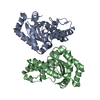
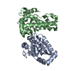
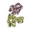
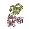
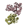
| Title | MANGANESE SUPEROXIDE DISMUTASE FROM ESCHERICHIA COLI | ||||||
 Components Components | MANGANESE SUPEROXIDE DISMUTASE | ||||||
 Keywords Keywords | OXIDOREDUCTASE / SUPEROXIDE DISMUTASE / MANGANESE ENZYME / METALLOPROTEIN / DNA BINDING | ||||||
| Function / homology |  Function and homology information Function and homology informationcellular response to selenium ion / response to acidic pH / superoxide metabolic process / superoxide dismutase / superoxide dismutase activity / antioxidant activity / removal of superoxide radicals / manganese ion binding / response to heat / response to oxidative stress ...cellular response to selenium ion / response to acidic pH / superoxide metabolic process / superoxide dismutase / superoxide dismutase activity / antioxidant activity / removal of superoxide radicals / manganese ion binding / response to heat / response to oxidative stress / protein homodimerization activity / DNA binding / metal ion binding / cytoplasm / cytosol Similarity search - Function | ||||||
| Biological species |  | ||||||
| Method |  X-RAY DIFFRACTION / MOLECULAR REPLACEMENT / Resolution: 2.1 Å X-RAY DIFFRACTION / MOLECULAR REPLACEMENT / Resolution: 2.1 Å | ||||||
 Authors Authors | Edwards, R.A. / Baker, H.M. / Whittaker, M.M. / Whittaker, J.W. / Jameson, G.B. / Baker, E.N. | ||||||
 Citation Citation | Journal: J.Biol.Inorg.Chem. / Year: 1998 Title: Crystal structure of Escherichia coli manganese superoxide dismutase at 2.1-angstrom resolution. Authors: Edwards, R.A. / Baker, H.M. / Whittaker, M.M. / Whittaker, J.W. / Jameson, G.B. / Baker, E.N. | ||||||
| History |
|
- Structure visualization
| Structure viewer | Molecule: MolmilJmol/JSmol |
|---|
- Downloads & links
-Download
| PDBx/mmCIF format | 1vew.cif.gz | 177.6 KB | Display | PDBx/mmCIF format |
|---|---|---|---|---|
| PDB format | pdb1vew.ent.gz | 142.5 KB | Display | PDB format |
| PDBx/mmJSON format | 1vew.json.gz | Tree view | PDBx/mmJSON format | |
| Others |  Other downloads Other downloads |
-Validation report
| Arichive directory | https://data.pdbj.org/pub/pdb/validation_reports/ve/1vewftp://data.pdbj.org/pub/pdb/validation_reports/ve/1vew | HTTPS FTP |
|---|
-Related structure data
| Related structure data |  1mngS S: Starting model for refinement |
|---|---|
| Similar structure data |


-Links
 PDBj
PDBj


- Assembly
| Deposited unit | 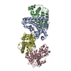
| ||||||||||||||||
|---|---|---|---|---|---|---|---|---|---|---|---|---|---|---|---|---|---|
| 1 | 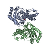
| ||||||||||||||||
| 2 | 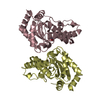
| ||||||||||||||||
| Unit cell |
| ||||||||||||||||
| Components on special symmetry positions |
| ||||||||||||||||
| Noncrystallographic symmetry (NCS) | NCS oper:
|
-Components
| #1: Protein | Mass: 22996.877 Da / Num. of mol.: 4 / Source method: isolated from a natural source / Source: (natural) #2: Chemical | ChemComp-MN /   Mass: 54.938 Da / Num. of mol.: 4 / Source method: obtained synthetically / Formula: Mn Mass: 54.938 Da / Num. of mol.: 4 / Source method: obtained synthetically / Formula: Mn#3: Chemical | ChemComp-OH /   Mass: 17.007 Da / Num. of mol.: 4 / Source method: obtained synthetically / Formula: HO Mass: 17.007 Da / Num. of mol.: 4 / Source method: obtained synthetically / Formula: HO#4: Water | ChemComp-HOH / |  Mass: 18.015 Da / Num. of mol.: 411 / Source method: isolated from a natural source / Formula: H2O Mass: 18.015 Da / Num. of mol.: 411 / Source method: isolated from a natural source / Formula: H2O |
|---|
-Experimental details
-Experiment
| Experiment | Method: X-RAY DIFFRACTION / Number of used crystals: 1 |
|---|
- Sample preparation
| Crystal | Density Matthews: 2.75 Å3/Da / Density % sol: 55 % | ||||||||||||||||||||||||||||||
|---|---|---|---|---|---|---|---|---|---|---|---|---|---|---|---|---|---|---|---|---|---|---|---|---|---|---|---|---|---|---|---|
| Crystal grow | Method: vapor diffusion, hanging drop / pH: 8.5 Details: CRYSTALS WERE GROWN AT ROOM TEMPERATURE USING THE HANGING DROP METHOD. THE HANGING DROPS WERE SUSPENDED OVER 0.75ML OF WELL SOLUTION (16-30% PEG6000 AND 0.05M BICINE TITRATED TO PH8.5) AND ...Details: CRYSTALS WERE GROWN AT ROOM TEMPERATURE USING THE HANGING DROP METHOD. THE HANGING DROPS WERE SUSPENDED OVER 0.75ML OF WELL SOLUTION (16-30% PEG6000 AND 0.05M BICINE TITRATED TO PH8.5) AND CONSISTED OF 2UL OF WELL SOLUTION AND 2UL OF PROTEIN SOLUTION (MNSOD AT 12MG/ML IN WATER)., vapor diffusion - hanging drop Temp details: room temp | ||||||||||||||||||||||||||||||
| Crystal grow | *PLUS Method: vapor diffusion, hanging drop | ||||||||||||||||||||||||||||||
| Components of the solutions | *PLUS
|
-Data collection
| Diffraction | Mean temperature: 293 K |
|---|---|
| Diffraction source | Source: ROTATING ANODE / Type: RIGAKU FR-C / Wavelength: 1.5418 |
| Detector | Type: RIGAKU / Detector: IMAGE PLATE / Date: Jul 1, 1996 / Details: 0.3MM DIAMETER COLLIMATOR |
| Radiation | Monochromator: GRAPHITE(002) / Monochromatic (M) / Laue (L): M / Scattering type: x-ray |
| Radiation wavelength | Wavelength: 1.5418 Å / Relative weight: 1 |
| Reflection | Resolution: 2.1→50 Å / Num. obs: 56890 / % possible obs: 96.8 % / Observed criterion σ(I): 0 / Redundancy: 3.7 % / Biso Wilson estimate: 23.3 Å2 / Rsym value: 0.056 / Net I/σ(I): 22 |
| Reflection shell | Resolution: 2.1→2.2 Å / Redundancy: 0.7 % / Mean I/σ(I) obs: 5.4 / Rsym value: 0.191 / % possible all: 77 |
| Reflection | *PLUS Num. measured all: 215051 / Rmerge(I) obs: 0.056 |
| Reflection shell | *PLUS % possible obs: 77 % / Rmerge(I) obs: 0.191 |
- Processing
| Software |
| ||||||||||||||||||||||||||||||||||||||||||||||||||
|---|---|---|---|---|---|---|---|---|---|---|---|---|---|---|---|---|---|---|---|---|---|---|---|---|---|---|---|---|---|---|---|---|---|---|---|---|---|---|---|---|---|---|---|---|---|---|---|---|---|---|---|
| Refinement | Method to determine structure: MOLECULAR REPLACEMENT Starting model: PDB ENTRY 1MNG Resolution: 2.1→50 Å / Isotropic thermal model: TNT BCORREL / σ(F): 0 / Stereochemistry target values: TNT CSDX-PROTGEO
| ||||||||||||||||||||||||||||||||||||||||||||||||||
| Solvent computation | Solvent model: MOEWS AND KRETSINGER (J.MOL.BIOL.(1975)91, 201-228) Bsol: 130 Å2 / ksol: 0.83 e/Å3 | ||||||||||||||||||||||||||||||||||||||||||||||||||
| Refinement step | Cycle: LAST / Resolution: 2.1→50 Å
| ||||||||||||||||||||||||||||||||||||||||||||||||||
| Refine LS restraints |
| ||||||||||||||||||||||||||||||||||||||||||||||||||
| Software | *PLUS Name: TNT / Version: 5E / Classification: refinement | ||||||||||||||||||||||||||||||||||||||||||||||||||
| Refinement | *PLUS Rfactor Rfree: 0.218 | ||||||||||||||||||||||||||||||||||||||||||||||||||
| Solvent computation | *PLUS | ||||||||||||||||||||||||||||||||||||||||||||||||||
| Displacement parameters | *PLUS | ||||||||||||||||||||||||||||||||||||||||||||||||||
| Refine LS restraints | *PLUS
|
