 Movie
Movie Controller

[English] 日本語
 Yorodumi
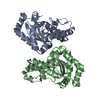
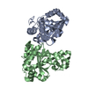
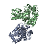
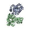
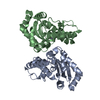
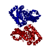
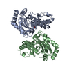
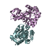
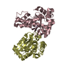
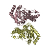
- PDB-1i0h: CRYSTAL STRUCTURE OF THE E. COLI MANGANESE SUPEROXIDE DISMUTASE M... -
+ Open data

- Basic information
| Entry | Database: PDB / ID: 1i0h | ||||||
|---|---|---|---|---|---|---|---|
| Title | CRYSTAL STRUCTURE OF THE E. COLI MANGANESE SUPEROXIDE DISMUTASE MUTANT Y174F AT 1.35 ANGSTROMS RESOLUTION. | ||||||
 Components Components | MANGANESE SUPEROXIDE DISMUTASE Y174F MUTANT | ||||||
 Keywords Keywords | OXIDOREDUCTASE / manganese superoxide dismutase / Y174F mutant / hydrogen bond / reactivity | ||||||
| Function / homology |  Function and homology information Function and homology informationcellular response to selenium ion / response to acidic pH / superoxide metabolic process / superoxide dismutase / superoxide dismutase activity / antioxidant activity / removal of superoxide radicals / manganese ion binding / response to heat / response to oxidative stress ...cellular response to selenium ion / response to acidic pH / superoxide metabolic process / superoxide dismutase / superoxide dismutase activity / antioxidant activity / removal of superoxide radicals / manganese ion binding / response to heat / response to oxidative stress / protein homodimerization activity / DNA binding / metal ion binding / cytosol / cytoplasm Similarity search - Function | ||||||
| Biological species |  | ||||||
| Method |  X-RAY DIFFRACTION / SYNCHROTRON / MOLECULAR REPLACEMENT / Resolution: 1.35 Å X-RAY DIFFRACTION / SYNCHROTRON / MOLECULAR REPLACEMENT / Resolution: 1.35 Å | ||||||
 Authors Authors | Edwards, R.A. / Whittaker, M.M. / Whittaker, J.W. / Baker, E.N. / Jameson, G.B. | ||||||
 Citation Citation | Journal: Biochemistry / Year: 2001 Title: Removing a hydrogen bond in the dimer interface of Escherichia coli manganese superoxide dismutase alters structure and reactivity. Authors: Edwards, R.A. / Whittaker, M.M. / Whittaker, J.W. / Baker, E.N. / Jameson, G.B. #1: Journal: Biochemistry / Year: 2001Title: Outer sphere mutations perturb metal reactivity in manganese superoxide dismutase Authors: Edwards, R.A. / Whittaker, M.M. / Whittaker, J.W. / Baker, E.N. / Jameson, G.B. | ||||||
| History |
|
- Structure visualization
| Structure viewer | Molecule: MolmilJmol/JSmol |
|---|
- Downloads & links
-Download
| PDBx/mmCIF format | 1i0h.cif.gz | 114.3 KB | Display | PDBx/mmCIF format |
|---|---|---|---|---|
| PDB format | pdb1i0h.ent.gz | 88 KB | Display | PDB format |
| PDBx/mmJSON format | 1i0h.json.gz | Tree view | PDBx/mmJSON format | |
| Others |  Other downloads Other downloads |
-Validation report
| Arichive directory | https://data.pdbj.org/pub/pdb/validation_reports/i0/1i0hftp://data.pdbj.org/pub/pdb/validation_reports/i0/1i0h | HTTPS FTP |
|---|
-Related structure data


-Links
 PDBj
PDBj
- Assembly
| Deposited unit | 
| ||||||||
|---|---|---|---|---|---|---|---|---|---|
| 1 |
| ||||||||
| Unit cell |
|
-Components
| #1: Protein | Mass: 22980.877 Da / Num. of mol.: 2 / Mutation: Y174F Source method: isolated from a genetically manipulated source Source: (gene. exp.) #2: Chemical |   Mass: 54.938 Da / Num. of mol.: 2 / Source method: obtained synthetically / Formula: Mn Mass: 54.938 Da / Num. of mol.: 2 / Source method: obtained synthetically / Formula: Mn#3: Water | ChemComp-HOH / |  Mass: 18.015 Da / Num. of mol.: 500 / Source method: isolated from a natural source / Formula: H2O Mass: 18.015 Da / Num. of mol.: 500 / Source method: isolated from a natural source / Formula: H2O |
|---|
-Experimental details
-Experiment
| Experiment | Method: X-RAY DIFFRACTION / Number of used crystals: 1 |
|---|
- Sample preparation
| Crystal | Density Matthews: 2.23 Å3/Da / Density % sol: 40 % | |||||||||||||||||||||||||
|---|---|---|---|---|---|---|---|---|---|---|---|---|---|---|---|---|---|---|---|---|---|---|---|---|---|---|
| Crystal grow | Temperature: 298 K / Method: vapor diffusion, sitting drop / pH: 8.5 Details: 16-20% PEG 6000, 0.1M bicine ph 8.5, VAPOR DIFFUSION, SITTING DROP, temperature 298K | |||||||||||||||||||||||||
| Crystal grow | *PLUS Details: drop consists of 2 micro litter of protein solution in 0.1 M bicine plus 2 micro litter of reservoir solutionPH range low: 8.3 / PH range high: 8 | |||||||||||||||||||||||||
| Components of the solutions | *PLUS
|
-Data collection
| Diffraction | Mean temperature: 110 K |
|---|---|
| Diffraction source | Source: SYNCHROTRON / Site: EMBL/DESY, HAMBURG  / Beamline: X11 / Wavelength: 0.9058 Å / Beamline: X11 / Wavelength: 0.9058 Å |
| Detector | Type: MARRESEARCH / Detector: AREA DETECTOR / Date: Nov 1, 1998 |
| Radiation | Monochromator: single-crystal germanium triangular monochromator Protocol: SINGLE WAVELENGTH / Monochromatic (M) / Laue (L): M / Scattering type: x-ray |
| Radiation wavelength | Wavelength: 0.9058 Å / Relative weight: 1 |
| Reflection | Resolution: 1.35→50 Å / Num. all: 89109 / Num. obs: 89109 / % possible obs: 100 % / Observed criterion σ(F): 0 / Observed criterion σ(I): 0 / Redundancy: 4.2 % / Biso Wilson estimate: 6.5 Å2 / Rmerge(I) obs: 0.027 / Net I/σ(I): 52.5 |
| Reflection shell | Resolution: 1.35→1.37 Å / Redundancy: 4.2 % / Rmerge(I) obs: 0.071 / Mean I/σ(I) obs: 18.3 / % possible all: 99.9 |
| Reflection | *PLUS Num. measured all: 561459 |
| Reflection shell | *PLUS % possible obs: 99.9 % |
- Processing
| Software |
| |||||||||||||||||||||||||||
|---|---|---|---|---|---|---|---|---|---|---|---|---|---|---|---|---|---|---|---|---|---|---|---|---|---|---|---|---|
| Refinement | Method to determine structure: MOLECULAR REPLACEMENT / Resolution: 1.35→50 Å / Num. parameters: 17529 / Num. restraintsaints: 16393 / σ(F): 4 / Stereochemistry target values: Engh & Huber
| |||||||||||||||||||||||||||
| Solvent computation | Solvent model: Moews & Kretsinger / Bsol: 2.19461 Å2 / ksol: 0.76955 e/Å3 | |||||||||||||||||||||||||||
| Refine analyze | Num. disordered residues: 0 / Occupancy sum hydrogen: 0 / Occupancy sum non hydrogen: 3667 | |||||||||||||||||||||||||||
| Refinement step | Cycle: LAST / Resolution: 1.35→50 Å
| |||||||||||||||||||||||||||
| Refine LS restraints |
|
