 Movie
Movie Controller

[English] 日本語
 Yorodumi
- PDB-1iu8: The X-ray Crystal Structure of Pyrrolidone-Carboxylate Peptidase ... -
+ Open data

- Basic information
| Entry | Database: PDB / ID: 1iu8 | ||||||
|---|---|---|---|---|---|---|---|
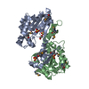
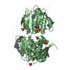
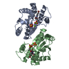
| Title | The X-ray Crystal Structure of Pyrrolidone-Carboxylate Peptidase from Hyperthermophilic Archaeon Pyrococcus horikoshii | ||||||
 Components Components | Pyrrolidone-carboxylate peptidase | ||||||
 Keywords Keywords | HYDROLASE / Thiol protease | ||||||
| Function / homology |  Function and homology information Function and homology informationpyroglutamyl-peptidase I / pyroglutamyl-peptidase activity / proteolysis / cytosol Similarity search - Function | ||||||
| Biological species |   Pyrococcus horikoshii (archaea) Pyrococcus horikoshii (archaea) | ||||||
| Method |  X-RAY DIFFRACTION / SYNCHROTRON / MOLECULAR REPLACEMENT / Resolution: 1.6 Å X-RAY DIFFRACTION / SYNCHROTRON / MOLECULAR REPLACEMENT / Resolution: 1.6 Å | ||||||
 Authors Authors | Sokabe, M. / Kawamura, T. / Sakai, N. / Yao, M. / Watanabe, N. / Tanaka, I. | ||||||
 Citation Citation | Journal: J.STRUCT.FUNCT.GENOM. / Year: 2002 Title: The X-ray crystal structure of pyrrolidone-carboxylate peptidase from hyperthermophilic archaea Pyrococcus horikoshii Authors: Sokabe, M. / Kawamura, T. / Sakai, N. / Yao, M. / Watanabe, N. / Tanaka, I. | ||||||
| History |
|
- Structure visualization
| Structure viewer | Molecule: MolmilJmol/JSmol |
|---|
- Downloads & links
-Download
| PDBx/mmCIF format | 1iu8.cif.gz | 99.5 KB | Display | PDBx/mmCIF format |
|---|---|---|---|---|
| PDB format | pdb1iu8.ent.gz | 77.2 KB | Display | PDB format |
| PDBx/mmJSON format | 1iu8.json.gz | Tree view | PDBx/mmJSON format | |
| Others |  Other downloads Other downloads |
-Validation report
| Arichive directory | https://data.pdbj.org/pub/pdb/validation_reports/iu/1iu8ftp://data.pdbj.org/pub/pdb/validation_reports/iu/1iu8 | HTTPS FTP |
|---|
-Related structure data
| Related structure data | 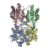 1iofS S: Starting model for refinement |
|---|---|
| Similar structure data |


-Links
 PDBj
PDBj- Assembly
| Deposited unit | 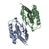
| ||||||||
|---|---|---|---|---|---|---|---|---|---|
| 1 |
| ||||||||
| 2 | 
| ||||||||
| Unit cell |
|
-Components
| #1: Protein | Mass: 22667.129 Da / Num. of mol.: 2 Source method: isolated from a genetically manipulated source Source: (gene. exp.) Pyrococcus horikoshii (archaea) / Gene: PH0596 / Plasmid: pET-22b(+) / Production host:  #2: Water | ChemComp-HOH / |  Mass: 18.015 Da / Num. of mol.: 501 / Source method: isolated from a natural source / Formula: H2O Mass: 18.015 Da / Num. of mol.: 501 / Source method: isolated from a natural source / Formula: H2O |
|---|
-Experimental details
-Experiment
| Experiment | Method: X-RAY DIFFRACTION / Number of used crystals: 1 |
|---|
- Sample preparation
| Crystal | Density Matthews: 2.3 Å3/Da / Density % sol: 53.5 % | |||||||||||||||||||||||||||||||||||
|---|---|---|---|---|---|---|---|---|---|---|---|---|---|---|---|---|---|---|---|---|---|---|---|---|---|---|---|---|---|---|---|---|---|---|---|---|
| Crystal grow | Temperature: 277 K / Method: vapor diffusion, hanging drop / pH: 7.4 Details: MPD, Magnesium Chlolide, Imidazole, pH 7.4, VAPOR DIFFUSION, HANGING DROP, temperature 277K | |||||||||||||||||||||||||||||||||||
| Crystal grow | *PLUS Temperature: 4 ℃ / PH range low: 9 / PH range high: 7.4 | |||||||||||||||||||||||||||||||||||
| Components of the solutions | *PLUS
|
-Data collection
| Diffraction | Mean temperature: 100 K |
|---|---|
| Diffraction source | Source: SYNCHROTRON / Site: Photon Factory  / Beamline: BL-18B / Wavelength: 1 Å / Beamline: BL-18B / Wavelength: 1 Å |
| Detector | Type: ADSC QUANTUM 4 / Detector: CCD / Date: Jun 23, 2001 / Details: mirrors |
| Radiation | Monochromator: Si(111) / Protocol: SINGLE WAVELENGTH / Monochromatic (M) / Laue (L): M / Scattering type: x-ray |
| Radiation wavelength | Wavelength: 1 Å / Relative weight: 1 |
| Reflection | Resolution: 1.6→23.6 Å / Num. all: 54101 / Num. obs: 54101 / % possible obs: 100 % / Observed criterion σ(I): 3 / Redundancy: 3.8 % / Biso Wilson estimate: 21.211 Å2 / Rmerge(I) obs: 0.052 / Rsym value: 0.052 / Net I/σ(I): 8 |
| Reflection shell | Resolution: 1.6→1.686 Å / Redundancy: 3.8 % / Rmerge(I) obs: 0.231 / Mean I/σ(I) obs: 3 / Num. unique all: 7861 / Rsym value: 0.231 / % possible all: 100 |
| Reflection | *PLUS Highest resolution: 1.6 Å / % possible obs: 100 % / Num. measured all: 207328 / Rmerge(I) obs: 0.061 |
| Reflection shell | *PLUS % possible obs: 100 % / Rmerge(I) obs: 0.269 / Mean I/σ(I) obs: 3.8 |
- Processing
| Software |
| ||||||||||||||||||||||||||||||||||||
|---|---|---|---|---|---|---|---|---|---|---|---|---|---|---|---|---|---|---|---|---|---|---|---|---|---|---|---|---|---|---|---|---|---|---|---|---|---|
| Refinement | Method to determine structure: MOLECULAR REPLACEMENT Starting model: PDB ENTRY 1IOF Resolution: 1.6→20 Å / Data cutoff high rms absF: 10000 / Isotropic thermal model: isotropic / Cross valid method: THROUGHOUT / σ(F): 0 / Stereochemistry target values: Engh & Huber
| ||||||||||||||||||||||||||||||||||||
| Solvent computation | Solvent model: throughout / Bsol: 49.57 Å2 / ksol: 0.36 e/Å3 | ||||||||||||||||||||||||||||||||||||
| Displacement parameters | Biso mean: 17.3 Å2
| ||||||||||||||||||||||||||||||||||||
| Refine analyze |
| ||||||||||||||||||||||||||||||||||||
| Refinement step | Cycle: LAST / Resolution: 1.6→20 Å
| ||||||||||||||||||||||||||||||||||||
| Refine LS restraints |
| ||||||||||||||||||||||||||||||||||||
| LS refinement shell | Resolution: 1.6→1.66 Å / Total num. of bins used: 10
| ||||||||||||||||||||||||||||||||||||
| Xplor file | Serial no: 1 / Param file: PROTEIN_REP.PARAM / Topol file: PROTEIN.TOP | ||||||||||||||||||||||||||||||||||||
| Refinement | *PLUS Rfactor obs: 0.189 | ||||||||||||||||||||||||||||||||||||
| Solvent computation | *PLUS | ||||||||||||||||||||||||||||||||||||
| Displacement parameters | *PLUS Biso mean: 17.3 Å2 | ||||||||||||||||||||||||||||||||||||
| Refine LS restraints | *PLUS
| ||||||||||||||||||||||||||||||||||||
| LS refinement shell | *PLUS Rfactor obs: 0.2689 |
