 Movie
Movie Controller


+ Open data

- Basic information
| Entry | Database: PDB / ID: 1il1 | ||||||
|---|---|---|---|---|---|---|---|
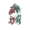
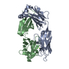
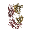
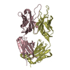
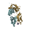
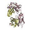
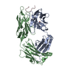
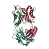
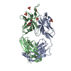
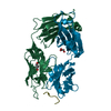
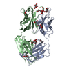
| Title | Crystal structure of G3-519, an anti-HIV monoclonal antibody | ||||||
 Components Components |
| ||||||
 Keywords Keywords | IMMUNE SYSTEM / Fab / beta sheet structure / antibody | ||||||
| Function / homology | Immunoglobulins / Immunoglobulin-like / Sandwich / Mainly Beta Function and homology information Function and homology information | ||||||
| Biological species |  | ||||||
| Method |  X-RAY DIFFRACTION / MOLECULAR REPLACEMENT / Resolution: 2.2 Å X-RAY DIFFRACTION / MOLECULAR REPLACEMENT / Resolution: 2.2 Å | ||||||
 Authors Authors | Berry, M.B. / Johnson, K.A. / Radding, W. / Fung, M. / Liou, R. / Phillips Jr., G.N. | ||||||
 Citation Citation | Journal: Proteins / Year: 2001 Title: Structure of an anti-HIV monoclonal Fab antibody fragment specific to a gp120 C-4 region peptide. Authors: Berry, M.B. / Johnson, K.A. / Radding, W. / Fung, M. / Liou, R. / Phillips Jr., G.N. | ||||||
| History |
|
- Structure visualization
| Structure viewer | Molecule: MolmilJmol/JSmol |
|---|
- Downloads & links
-Download
| PDBx/mmCIF format | 1il1.cif.gz | 102 KB | Display | PDBx/mmCIF format |
|---|---|---|---|---|
| PDB format | pdb1il1.ent.gz | 78 KB | Display | PDB format |
| PDBx/mmJSON format | 1il1.json.gz | Tree view | PDBx/mmJSON format | |
| Others |  Other downloads Other downloads |
-Validation report
| Summary document | 1il1_validation.pdf.gz | 432.8 KB | Display | wwPDB validaton report |
|---|---|---|---|---|
| Full document | 1il1_full_validation.pdf.gz | 441.7 KB | Display | |
| Data in XML | 1il1_validation.xml.gz | 21.2 KB | Display | |
| Data in CIF | 1il1_validation.cif.gz | 30.1 KB | Display | |
| Arichive directory | https://data.pdbj.org/pub/pdb/validation_reports/il/1il1ftp://data.pdbj.org/pub/pdb/validation_reports/il/1il1 | HTTPS FTP |
-Related structure data
| Similar structure data |
|---|


-Links
 PDBj
PDBj

- Assembly
| Deposited unit | 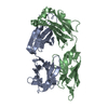
| ||||||||
|---|---|---|---|---|---|---|---|---|---|
| 1 |
| ||||||||
| Unit cell |
|
-Components
| #1: Antibody | Mass: 23559.076 Da / Num. of mol.: 1 / Fragment: FAB FRAGMENT / Source method: isolated from a natural source / Source: (natural) Cell line: mouse myeloma line SP2/0 fused with antibody expressing B cells |
|---|---|
| #2: Antibody | Mass: 24514.133 Da / Num. of mol.: 1 / Fragment: FAB FRAGMENT / Source method: isolated from a natural source / Source: (natural) Cell line: mouse myeloma line SP2/0 fused with antibody expressing B cells |
| #3: Water | ChemComp-HOH /  Mass: 18.015 Da / Num. of mol.: 260 / Source method: isolated from a natural source / Formula: H2O Mass: 18.015 Da / Num. of mol.: 260 / Source method: isolated from a natural source / Formula: H2O |
| Has protein modification | Y |
-Experimental details
-Experiment
| Experiment | Method: X-RAY DIFFRACTION / Number of used crystals: 1 |
|---|
- Sample preparation
| Crystal | Density Matthews: 2.18 Å3/Da / Density % sol: 43.69 % | ||||||||||||||||||||||||
|---|---|---|---|---|---|---|---|---|---|---|---|---|---|---|---|---|---|---|---|---|---|---|---|---|---|
| Crystal grow | Temperature: 296 K / Method: vapor diffusion, hanging drop / pH: 5.5 Details: 2M ammonium sulfate, 200mM sodium chloride, 100mM sodium citrate, pH 5.5, VAPOR DIFFUSION, HANGING DROP, temperature 296K | ||||||||||||||||||||||||
| Crystal grow | *PLUS Details: used microseeding / PH range low: 6 / PH range high: 5 | ||||||||||||||||||||||||
| Components of the solutions | *PLUS
|
-Data collection
| Diffraction | Mean temperature: 296 K |
|---|---|
| Diffraction source | Source: ROTATING ANODE / Type: SIEMENS / Wavelength: 1.5418 |
| Detector | Type: RIGAKU RAXIS II / Detector: IMAGE PLATE / Date: Apr 22, 1997 |
| Radiation | Monochromator: graphite / Protocol: SINGLE WAVELENGTH / Monochromatic (M) / Laue (L): M / Scattering type: x-ray |
| Radiation wavelength | Wavelength: 1.5418 Å / Relative weight: 1 |
| Reflection | Resolution: 2.2→19.74 Å / Num. all: 18605 / Num. obs: 18605 / % possible obs: 90.2 % / Observed criterion σ(F): 0 / Observed criterion σ(I): 0 / Redundancy: 3 % / Biso Wilson estimate: 18.7 Å2 / Rmerge(I) obs: 0.1 / Net I/σ(I): 12.4 |
| Reflection shell | Resolution: 2.2→2.28 Å / Redundancy: 1.9 % / Rmerge(I) obs: 0.505 / % possible all: 88.1 |
| Reflection | *PLUS |
- Processing
| Software |
| ||||||||||||||||||||||||||||||||||||||||
|---|---|---|---|---|---|---|---|---|---|---|---|---|---|---|---|---|---|---|---|---|---|---|---|---|---|---|---|---|---|---|---|---|---|---|---|---|---|---|---|---|---|
| Refinement | Method to determine structure: MOLECULAR REPLACEMENT / Resolution: 2.2→19.74 Å / Rfactor Rfree error: 0.006 / Data cutoff high absF: 124012.54 / Data cutoff low absF: 0 / Isotropic thermal model: RESTRAINED / Cross valid method: THROUGHOUT / σ(F): 0 / σ(I): 0
| ||||||||||||||||||||||||||||||||||||||||
| Solvent computation | Solvent model: FLAT MODEL / Bsol: 60.51 Å2 / ksol: 0.345 e/Å3 | ||||||||||||||||||||||||||||||||||||||||
| Displacement parameters | Biso mean: 35.8 Å2
| ||||||||||||||||||||||||||||||||||||||||
| Refine analyze |
| ||||||||||||||||||||||||||||||||||||||||
| Refinement step | Cycle: LAST / Resolution: 2.2→19.74 Å
| ||||||||||||||||||||||||||||||||||||||||
| Refine LS restraints |
| ||||||||||||||||||||||||||||||||||||||||
| LS refinement shell | Resolution: 2.19→2.33 Å / Rfactor Rfree error: 0.022 / Total num. of bins used: 6
| ||||||||||||||||||||||||||||||||||||||||
| Xplor file |
| ||||||||||||||||||||||||||||||||||||||||
| Software | *PLUS Name: CNS / Version: 1 / Classification: refinement | ||||||||||||||||||||||||||||||||||||||||
| Refinement | *PLUS σ(F): 0 / % reflection Rfree: 9.9 % / Rfactor Rfree: 0.247 | ||||||||||||||||||||||||||||||||||||||||
| Solvent computation | *PLUS | ||||||||||||||||||||||||||||||||||||||||
| Displacement parameters | *PLUS Biso mean: 35.8 Å2 | ||||||||||||||||||||||||||||||||||||||||
| Refine LS restraints | *PLUS
| ||||||||||||||||||||||||||||||||||||||||
| LS refinement shell | *PLUS Rfactor Rfree: 0.351 / % reflection Rfree: 9.9 % / Rfactor Rwork: 0.288 |
