 Movie
Movie Controller

[English] 日本語
 Yorodumi
- PDB-1ig4: Solution Structure of the Methyl-CpG-Binding Domain of Human MBD1... -
+ Open data

- Basic information
| Entry | Database: PDB / ID: 1ig4 | ||||||
|---|---|---|---|---|---|---|---|
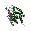
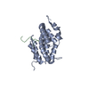
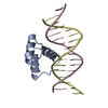
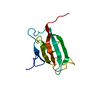
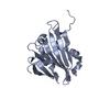
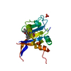
| Title | Solution Structure of the Methyl-CpG-Binding Domain of Human MBD1 in Complex with Methylated DNA | ||||||
 Components Components |
| ||||||
 Keywords Keywords | TRANSCRIPTION/DNA / PROTEIN-DNA COMPLEX / ALPHA-BETA / DOUBLE HELIX / RECOGNITION VIA BETA-SHEET / TRANSCRIPTION-DNA COMPLEX | ||||||
| Function / homology |  Function and homology information Function and homology informationdouble-stranded methylated DNA binding / unmethylated CpG binding / methyl-CpG binding / DNA methylation-dependent constitutive heterochromatin formation / SUMOylation of transcription cofactors / nuclear matrix / chromosome / transcription by RNA polymerase II / nuclear speck / negative regulation of DNA-templated transcription ...double-stranded methylated DNA binding / unmethylated CpG binding / methyl-CpG binding / DNA methylation-dependent constitutive heterochromatin formation / SUMOylation of transcription cofactors / nuclear matrix / chromosome / transcription by RNA polymerase II / nuclear speck / negative regulation of DNA-templated transcription / negative regulation of transcription by RNA polymerase II / DNA binding / zinc ion binding / nucleoplasm / nucleus Similarity search - Function | ||||||
| Biological species |  Homo sapiens (human) Homo sapiens (human) | ||||||
| Method | SOLUTION NMR / Structure calculations were performed following simulated annealing protocols using X-PLOR. | ||||||
 Authors Authors | Ohki, I. / Shimotake, N. / Fujita, N. / Jee, J.-G. / Ikegami, T. / Nakao, M. / Shirakawa, M. | ||||||
 Citation Citation | Journal: Cell(Cambridge,Mass.) / Year: 2001 Title: Solution structure of the methyl-CpG binding domain of human MBD1 in complex with methylated DNA. Authors: Ohki, I. / Shimotake, N. / Fujita, N. / Jee, J. / Ikegami, T. / Nakao, M. / Shirakawa, M. #1: Journal: Embo J. / Year: 1999Title: Solution structure of the methyl-CpG-binding domain of the methylation-dependent transcriptional repressor MBD1 Authors: Ohki, I. / Shimotake, N. / Fujita, N. / Nakao, M. / Shirakawa, M. #2: Journal: Nat.Genet. / Year: 1997Title: A component of the transcriptional repressor MeCP1 shares a motif with DNA methyltransferase and HRX proteins Authors: Cross, S.H. / Meehan, R.R. / Nan, X. / Bird, A. | ||||||
| History |
|
- Structure visualization
| Structure viewer | Molecule: MolmilJmol/JSmol |
|---|
- Downloads & links
-Download
| PDBx/mmCIF format | 1ig4.cif.gz | 777.1 KB | Display | PDBx/mmCIF format |
|---|---|---|---|---|
| PDB format | pdb1ig4.ent.gz | 638.1 KB | Display | PDB format |
| PDBx/mmJSON format | 1ig4.json.gz | Tree view | PDBx/mmJSON format | |
| Others |  Other downloads Other downloads |
-Validation report
| Arichive directory | https://data.pdbj.org/pub/pdb/validation_reports/ig/1ig4ftp://data.pdbj.org/pub/pdb/validation_reports/ig/1ig4 | HTTPS FTP |
|---|
-Related structure data
| Similar structure data |
|---|


-Links
 PDBj
PDBj


- Assembly
| Deposited unit | 
| |||||||||
|---|---|---|---|---|---|---|---|---|---|---|
| 1 |
| |||||||||
| NMR ensembles |
|
-Components
| #1: DNA chain | Mass: 3676.431 Da / Num. of mol.: 2 / Source method: obtained synthetically #2: Protein | | Mass: 8516.751 Da / Num. of mol.: 1 / Fragment: Methyl-CpG-binding domain Source method: isolated from a genetically manipulated source Source: (gene. exp.) Homo sapiens (human) / Gene: MBD1 / Plasmid: pGEX-2T / Production host:  |
|---|
-Experimental details
-Experiment
| Experiment | Method: SOLUTION NMR | ||||||||||||||||||||||||
|---|---|---|---|---|---|---|---|---|---|---|---|---|---|---|---|---|---|---|---|---|---|---|---|---|---|
| NMR experiment |
| ||||||||||||||||||||||||
| NMR details | Text: The structure was determined using multi-dimensional heteronuclear techniques. |
- Sample preparation
| Details |
| ||||||||||||
|---|---|---|---|---|---|---|---|---|---|---|---|---|---|
| Sample conditions | pH: 6.5 / Temperature: 303 K | ||||||||||||
| Crystal grow | *PLUS Method: other / Details: NMR |
-NMR measurement
| NMR spectrometer |
|
|---|
- Processing
| NMR software |
| ||||||||||||||||||||
|---|---|---|---|---|---|---|---|---|---|---|---|---|---|---|---|---|---|---|---|---|---|
| Refinement | Method: Structure calculations were performed following simulated annealing protocols using X-PLOR. Software ordinal: 1 Details: The structure was determined from a total of 2,022 distance and dihedral angle restraints, including 91 intermolecular NOE restraints. | ||||||||||||||||||||
| NMR representative | Selection criteria: lowest energy | ||||||||||||||||||||
| NMR ensemble | Conformer selection criteria: structures with the lowest energy Conformers calculated total number: 200 / Conformers submitted total number: 20 |
 NMRPipe