| Entry | Database: PDB / ID: 4rth
|
|---|
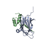
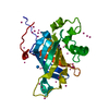
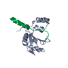
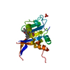
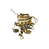
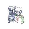
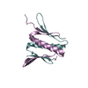
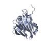
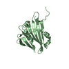
| Title | The crystal structure of PsbP from Zea mays |
|---|
 Components Components | Membrane-extrinsic protein of photosystem II PsbP |
|---|
 Keywords Keywords | PHOTOSYNTHESIS / beta-sandwich folding / membrane-extrinsic protein of photosystem II |
|---|
| Function / homology | Mog1/PsbP, alpha/beta/alpha sandwich / Protein Transport Mog1p; Chain A / 3-Layer(aba) Sandwich / Alpha Beta / :  Function and homology information Function and homology information |
|---|
| Biological species |   Zea mays (maize) Zea mays (maize) |
|---|
| Method |  X-RAY DIFFRACTION / SYNCHROTRON / MOLECULAR REPLACEMENT / Resolution: 1.6 Å X-RAY DIFFRACTION / SYNCHROTRON / MOLECULAR REPLACEMENT / Resolution: 1.6 Å |
|---|
 Authors Authors | Cao, P. / Xie, Y. / Li, M. / Pan, X.W. / Zhang, H.M. / Zhao, X.L. / Su, X.D. / Cheng, T. / Chang, W. |
|---|
 Citation Citation | Journal: Mol Plant / Year: 2015
Title: Crystal structure analysis of extrinsic PsbP protein of photosystem II reveals a manganese-induced conformational change.
Authors: Cao, P. / Xie, Y. / Li, M. / Pan, X. / Zhang, H. / Zhao, X. / Su, X. / Cheng, T. / Chang, W. |
|---|
| History | | Deposition | Nov 15, 2014 | Deposition site: RCSB / Processing site: PDBJ |
|---|
| Revision 1.0 | Mar 11, 2015 | Provider: repository / Type: Initial release |
|---|
| Revision 1.1 | Aug 24, 2022 | Group: Database references / Category: citation / citation_author / database_2
Item: _citation.journal_volume / _citation.page_first ..._citation.journal_volume / _citation.page_first / _citation.page_last / _citation.title / _citation_author.name / _database_2.pdbx_DOI / _database_2.pdbx_database_accession |
|---|
| Revision 1.2 | Nov 8, 2023 | Group: Data collection / Refinement description
Category: chem_comp_atom / chem_comp_bond ...chem_comp_atom / chem_comp_bond / pdbx_initial_refinement_model / struct_ncs_dom_lim
Item: _struct_ncs_dom_lim.beg_auth_comp_id / _struct_ncs_dom_lim.beg_label_asym_id ..._struct_ncs_dom_lim.beg_auth_comp_id / _struct_ncs_dom_lim.beg_label_asym_id / _struct_ncs_dom_lim.beg_label_comp_id / _struct_ncs_dom_lim.beg_label_seq_id / _struct_ncs_dom_lim.end_auth_comp_id / _struct_ncs_dom_lim.end_label_asym_id / _struct_ncs_dom_lim.end_label_comp_id / _struct_ncs_dom_lim.end_label_seq_id |
|---|
|
|---|
 Movie
Movie Controller


 Open data
 Basic information
 Structure visualization
 Downloads & links
 Other downloads


 PDBj
PDBj Assembly
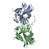
 Mass: 18.015 Da / Num. of mol.: 266 / Source method: isolated from a natural source / Formula: H2O
 Sample preparation
 / Beamline: AR-NW12A / Wavelength: 1.26 Å
 Processing