 Movie
Movie Controller

[English] 日本語
 Yorodumi
- PDB-1ifb: REFINED APOPROTEIN STRUCTURE OF RAT INTESTINAL FATTY ACID BINDING... -
+ Open data

- Basic information
| Entry | Database: PDB / ID: 1ifb | ||||||
|---|---|---|---|---|---|---|---|
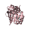
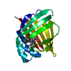
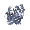
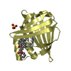
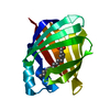
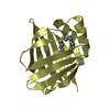
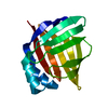
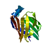
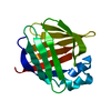
| Title | REFINED APOPROTEIN STRUCTURE OF RAT INTESTINAL FATTY ACID BINDING PROTEIN PRODUCED IN ESCHERICHIA COLI | ||||||
 Components Components | INTESTINAL FATTY ACID BINDING PROTEIN | ||||||
 Keywords Keywords | FATTY ACID-BINDING PROTEIN | ||||||
| Function / homology |  Function and homology information Function and homology informationTriglyceride catabolism / intestinal lipid absorption / triglyceride biosynthetic process / lipid droplet formation / apical cortex / intestinal absorption / response to food / long-chain fatty acid binding / long-chain fatty acid transmembrane transporter activity / response to starvation ...Triglyceride catabolism / intestinal lipid absorption / triglyceride biosynthetic process / lipid droplet formation / apical cortex / intestinal absorption / response to food / long-chain fatty acid binding / long-chain fatty acid transmembrane transporter activity / response to starvation / microvillus / long-chain fatty acid transport / fatty acid transport / energy homeostasis / fatty acid metabolic process / fatty acid binding / lipid metabolic process / nucleus / cytosol Similarity search - Function | ||||||
| Biological species |  | ||||||
| Method |  X-RAY DIFFRACTION / Resolution: 1.96 Å X-RAY DIFFRACTION / Resolution: 1.96 Å | ||||||
 Authors Authors | Sacchettini, J.C. / Gordon, J.I. / Banaszak, L.J. | ||||||
 Citation Citation | Journal: Proc.Natl.Acad.Sci.USA / Year: 1989 Title: Refined apoprotein structure of rat intestinal fatty acid binding protein produced in Escherichia coli. Authors: Sacchettini, J.C. / Gordon, J.I. / Banaszak, L.J. | ||||||
| History |
|
- Structure visualization
| Structure viewer | Molecule: MolmilJmol/JSmol |
|---|
- Downloads & links
-Download
| PDBx/mmCIF format | 1ifb.cif.gz | 37.7 KB | Display | PDBx/mmCIF format |
|---|---|---|---|---|
| PDB format | pdb1ifb.ent.gz | 26.2 KB | Display | PDB format |
| PDBx/mmJSON format | 1ifb.json.gz | Tree view | PDBx/mmJSON format | |
| Others |  Other downloads Other downloads |
-Validation report
| Arichive directory | https://data.pdbj.org/pub/pdb/validation_reports/if/1ifbftp://data.pdbj.org/pub/pdb/validation_reports/if/1ifb | HTTPS FTP |
|---|
-Related structure data
| Similar structure data |
|---|

-Links
 PDBj
PDBj

- Assembly
| Deposited unit | 
| ||||||||
|---|---|---|---|---|---|---|---|---|---|
| 1 |
| ||||||||
| Unit cell |
|
-Components
| #1: Protein | Mass: 15015.015 Da / Num. of mol.: 1 Source method: isolated from a genetically manipulated source Source: (gene. exp.) |
|---|---|
| #2: Water | ChemComp-HOH /  Mass: 18.015 Da / Num. of mol.: 52 / Source method: isolated from a natural source / Formula: H2O Mass: 18.015 Da / Num. of mol.: 52 / Source method: isolated from a natural source / Formula: H2O |
-Experimental details
-Experiment
| Experiment | Method: X-RAY DIFFRACTION |
|---|
- Sample preparation
| Crystal | Density Matthews: 1.97 Å3/Da / Density % sol: 37.63 % | ||||||||||||||||||||
|---|---|---|---|---|---|---|---|---|---|---|---|---|---|---|---|---|---|---|---|---|---|
| Crystal grow | *PLUS pH: 7.3 / Method: free interface diffusion | ||||||||||||||||||||
| Components of the solutions | *PLUS
|
-Data collection
| Radiation | Scattering type: x-ray |
|---|---|
| Radiation wavelength | Relative weight: 1 |
| Reflection | *PLUS Highest resolution: 2.5 Å / Lowest resolution: 10 Å / Rmerge(I) obs: 0.082 |
| Reflection shell | *PLUS Highest resolution: 2 Å / Lowest resolution: 2.5 Å / Rmerge(I) obs: 0.162 |
- Processing
| Software | Name: X-PLOR / Classification: refinement | ||||||||||||||||||||||||||||||||||||||||||||||||||||||||||||
|---|---|---|---|---|---|---|---|---|---|---|---|---|---|---|---|---|---|---|---|---|---|---|---|---|---|---|---|---|---|---|---|---|---|---|---|---|---|---|---|---|---|---|---|---|---|---|---|---|---|---|---|---|---|---|---|---|---|---|---|---|---|
| Refinement | Rfactor Rwork: 0.188 / Highest resolution: 1.96 Å | ||||||||||||||||||||||||||||||||||||||||||||||||||||||||||||
| Refinement step | Cycle: LAST / Highest resolution: 1.96 Å
| ||||||||||||||||||||||||||||||||||||||||||||||||||||||||||||
| Refine LS restraints |
| ||||||||||||||||||||||||||||||||||||||||||||||||||||||||||||
| Refinement | *PLUS Highest resolution: 1.96 Å / Lowest resolution: 7 Å / Num. reflection obs: 5990 / σ(F): 1.5 / Rfactor obs: 0.188 | ||||||||||||||||||||||||||||||||||||||||||||||||||||||||||||
| Solvent computation | *PLUS | ||||||||||||||||||||||||||||||||||||||||||||||||||||||||||||
| Displacement parameters | *PLUS | ||||||||||||||||||||||||||||||||||||||||||||||||||||||||||||
| Refine LS restraints | *PLUS Type: x_angle_d |
