 Movie
Movie Controller

[English] 日本語
 Yorodumi
- PDB-1idr: CRYSTAL STRUCTURE OF THE TRUNCATED-HEMOGLOBIN-N FROM MYCOBACTERIU... -
+ Open data

- Basic information
| Entry | Database: PDB / ID: 1idr | ||||||
|---|---|---|---|---|---|---|---|
| Title | CRYSTAL STRUCTURE OF THE TRUNCATED-HEMOGLOBIN-N FROM MYCOBACTERIUM TUBERCULOSIS | ||||||
 Components Components | HEMOGLOBIN HBN | ||||||
 Keywords Keywords | OXYGEN STORAGE/TRANSPORT / truncated hemoglobin fold / OXYGEN STORAGE-TRANSPORT COMPLEX | ||||||
| Function / homology |  Function and homology information Function and homology informationdetoxification of nitrogen compound / Tolerance by Mtb to nitric oxide produced by macrophages / nitric oxide dioxygenase [NAD(P)H] activity / nitric oxide catabolic process / thioredoxin peroxidase activity / cell redox homeostasis / oxygen carrier activity / oxygen binding / cellular response to oxidative stress / heme binding ...detoxification of nitrogen compound / Tolerance by Mtb to nitric oxide produced by macrophages / nitric oxide dioxygenase [NAD(P)H] activity / nitric oxide catabolic process / thioredoxin peroxidase activity / cell redox homeostasis / oxygen carrier activity / oxygen binding / cellular response to oxidative stress / heme binding / metal ion binding / cytoplasm / cytosol Similarity search - Function | ||||||
| Biological species |   Mycobacterium tuberculosis (bacteria) Mycobacterium tuberculosis (bacteria) | ||||||
| Method |  X-RAY DIFFRACTION / SYNCHROTRON / MOLECULAR REPLACEMENT / Resolution: 1.9 Å X-RAY DIFFRACTION / SYNCHROTRON / MOLECULAR REPLACEMENT / Resolution: 1.9 Å | ||||||
 Authors Authors | Milani, M. / Pesce, A. / Ascenzi, P. / Guertin, M. / Bolognesi, M. | ||||||
 Citation Citation | Journal: EMBO J. / Year: 2001 Title: Mycobacterium tuberculosis hemoglobin N displays a protein tunnel suited for O2 diffusion to the heme. Authors: Milani, M. / Pesce, A. / Ouellet, Y. / Ascenzi, P. / Guertin, M. / Bolognesi, M. | ||||||
| History |
| ||||||
| Remark 300 | BIOMOLECULE: 1 THIS ENTRY CONTAINS THE CRYSTALLOGRAPHIC ASYMMETRIC UNIT WHICH CONSISTS OF 2 CHAIN(S) ...BIOMOLECULE: 1 THIS ENTRY CONTAINS THE CRYSTALLOGRAPHIC ASYMMETRIC UNIT WHICH CONSISTS OF 2 CHAIN(S). SEE REMARK 350 FOR INFORMATION ON GENERATING THE BIOLOGICAL MOLECULE(S). Gel-filtration experiment indicates that the protein is loosely dimeric. |
- Structure visualization
| Structure viewer | Molecule: MolmilJmol/JSmol |
|---|
- Downloads & links
-Download
| PDBx/mmCIF format | 1idr.cif.gz | 68.6 KB | Display | PDBx/mmCIF format |
|---|---|---|---|---|
| PDB format | pdb1idr.ent.gz | 51.1 KB | Display | PDB format |
| PDBx/mmJSON format | 1idr.json.gz | Tree view | PDBx/mmJSON format | |
| Others |  Other downloads Other downloads |
-Validation report
| Arichive directory | https://data.pdbj.org/pub/pdb/validation_reports/id/1idrftp://data.pdbj.org/pub/pdb/validation_reports/id/1idr | HTTPS FTP |
|---|
-Related structure data
| Similar structure data |
|---|


-Links
 PDBj
PDBj


- Assembly
| Deposited unit | 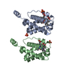
| ||||||||
|---|---|---|---|---|---|---|---|---|---|
| 1 |
| ||||||||
| Unit cell |
|
-Components
| #1: Protein | Mass: 14464.515 Da / Num. of mol.: 2 Source method: isolated from a genetically manipulated source Source: (gene. exp.) Mycobacterium tuberculosis (bacteria) / Production host: #2: Chemical |   Mass: 94.971 Da / Num. of mol.: 3 / Source method: obtained synthetically / Formula: PO4 Mass: 94.971 Da / Num. of mol.: 3 / Source method: obtained synthetically / Formula: PO4#3: Chemical |   Mass: 616.487 Da / Num. of mol.: 2 / Source method: obtained synthetically / Formula: C34H32FeN4O4 Mass: 616.487 Da / Num. of mol.: 2 / Source method: obtained synthetically / Formula: C34H32FeN4O4#4: Chemical |   Mass: 31.999 Da / Num. of mol.: 2 / Source method: obtained synthetically / Formula: O2 Mass: 31.999 Da / Num. of mol.: 2 / Source method: obtained synthetically / Formula: O2#5: Water | ChemComp-HOH / |  Mass: 18.015 Da / Num. of mol.: 228 / Source method: isolated from a natural source / Formula: H2O Mass: 18.015 Da / Num. of mol.: 228 / Source method: isolated from a natural source / Formula: H2O |
|---|
-Experimental details
-Experiment
| Experiment | Method: X-RAY DIFFRACTION / Number of used crystals: 1 |
|---|
- Sample preparation
| Crystal | Density Matthews: 2.19 Å3/Da / Density % sol: 43.9 % | ||||||||||||||||||
|---|---|---|---|---|---|---|---|---|---|---|---|---|---|---|---|---|---|---|---|
| Crystal grow | Temperature: 277 K / Method: microdialysis / pH: 8.3 Details: potassium phosphate, pH 8.3, MICRODIALYSIS, temperature 277K | ||||||||||||||||||
| Crystal grow | *PLUS Temperature: 4 ℃ | ||||||||||||||||||
| Components of the solutions | *PLUS
|
-Data collection
| Diffraction | Mean temperature: 100 K |
|---|---|
| Diffraction source | Source: SYNCHROTRON / Site: ESRF  / Beamline: ID14-3 / Wavelength: 0.931 Å / Beamline: ID14-3 / Wavelength: 0.931 Å |
| Detector | Type: MARRESEARCH / Detector: CCD / Date: Apr 27, 2000 |
| Radiation | Protocol: SINGLE WAVELENGTH / Monochromatic (M) / Laue (L): M / Scattering type: x-ray |
| Radiation wavelength | Wavelength: 0.931 Å / Relative weight: 1 |
| Reflection | Resolution: 1.9→40 Å / Num. all: 190583 / Num. obs: 18688 / % possible obs: 95 % / Observed criterion σ(F): 0 / Observed criterion σ(I): 2 / Redundancy: 10.2 % / Rmerge(I) obs: 0.053 / Net I/σ(I): 18.1 |
| Reflection shell | Resolution: 1.9→1.93 Å / Rmerge(I) obs: 0.331 / % possible all: 88.9 |
| Reflection | *PLUS Lowest resolution: 40 Å / % possible obs: 95.1 % / Redundancy: 3.5 % / Num. measured all: 190583 |
| Reflection shell | *PLUS % possible obs: 88.9 % / Redundancy: 2.3 % / Mean I/σ(I) obs: 3 |
- Processing
| Software |
| |||||||||||||||||||||||||
|---|---|---|---|---|---|---|---|---|---|---|---|---|---|---|---|---|---|---|---|---|---|---|---|---|---|---|
| Refinement | Method to determine structure: MOLECULAR REPLACEMENT / Resolution: 1.9→50 Å / SU B: 4.43152 / SU ML: 0.12857 / Cross valid method: THROUGHOUT / σ(F): 0 / σ(I): 0 / ESU R: 0.17577 / ESU R Free: 0.16852 / Stereochemistry target values: Engh and Huber
| |||||||||||||||||||||||||
| Displacement parameters | Biso mean: 32.725 Å2
| |||||||||||||||||||||||||
| Refine analyze | Luzzati sigma a obs: 0.04 Å | |||||||||||||||||||||||||
| Refinement step | Cycle: LAST / Resolution: 1.9→50 Å
| |||||||||||||||||||||||||
| Refine LS restraints |
| |||||||||||||||||||||||||
| Software | *PLUS Name: REFMAC / Classification: refinement | |||||||||||||||||||||||||
| Refinement | *PLUS Highest resolution: 1.9 Å / Lowest resolution: 50 Å / σ(F): 0 / % reflection Rfree: 5.2 % | |||||||||||||||||||||||||
| Solvent computation | *PLUS | |||||||||||||||||||||||||
| Displacement parameters | *PLUS | |||||||||||||||||||||||||
| Refine LS restraints | *PLUS
|
