 Movie
Movie Controller

[English] 日本語
 Yorodumi
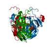
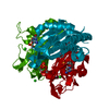
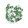
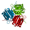
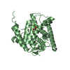
- PDB-2py4: Full length structure of the Mycobacterium tuberculosis dUTPase c... -
+ Open data

- Basic information
| Entry | Database: PDB / ID: 2py4 | ||||||
|---|---|---|---|---|---|---|---|
| Title | Full length structure of the Mycobacterium tuberculosis dUTPase complexed with magnesium and alpha,beta-imido-dUTP. | ||||||
 Components Components | Deoxyuridine 5'-triphosphate nucleotidohydrolase | ||||||
 Keywords Keywords | HYDROLASE / JELLY-ROLL / enzyme-ligand complex | ||||||
| Function / homology |  Function and homology information Function and homology informationdUTP metabolic process / dUTP catabolic process / dUMP biosynthetic process / dUTP diphosphatase / dUTP diphosphatase activity / magnesium ion binding Similarity search - Function | ||||||
| Biological species |   Mycobacterium tuberculosis (bacteria) Mycobacterium tuberculosis (bacteria) | ||||||
| Method |  X-RAY DIFFRACTION / MOLECULAR REPLACEMENT / Resolution: 1.49 Å X-RAY DIFFRACTION / MOLECULAR REPLACEMENT / Resolution: 1.49 Å | ||||||
 Authors Authors | Barabas, O. / Nagy, N. / Takacs, E. / Vertessy, B.G. | ||||||
 Citation Citation | Journal: Biochem.Biophys.Res.Commun. / Year: 2008 Title: Active site of mycobacterial dUTPase: structural characteristics and a built-in sensor. Authors: Varga, B. / Barabas, O. / Takacs, E. / Nagy, N. / Nagy, P. / Vertessy, B.G. | ||||||
| History |
|
- Structure visualization
| Structure viewer | Molecule: MolmilJmol/JSmol |
|---|
- Downloads & links
-Download
| PDBx/mmCIF format | 2py4.cif.gz | 48 KB | Display | PDBx/mmCIF format |
|---|---|---|---|---|
| PDB format | pdb2py4.ent.gz | 31.8 KB | Display | PDB format |
| PDBx/mmJSON format | 2py4.json.gz | Tree view | PDBx/mmJSON format | |
| Others |  Other downloads Other downloads |
-Validation report
| Arichive directory | https://data.pdbj.org/pub/pdb/validation_reports/py/2py4ftp://data.pdbj.org/pub/pdb/validation_reports/py/2py4 | HTTPS FTP |
|---|
-Related structure data
| Related structure data |  1mq7S S: Starting model for refinement |
|---|---|
| Similar structure data |


-Links
 PDBj
PDBj
- Assembly
| Deposited unit | 
| |||||||||||||||||||||
|---|---|---|---|---|---|---|---|---|---|---|---|---|---|---|---|---|---|---|---|---|---|---|
| 1 | 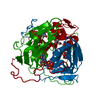
| |||||||||||||||||||||
| Unit cell |
| |||||||||||||||||||||
| Components on special symmetry positions |
| |||||||||||||||||||||
| Details | The biological assembly is a trimer generated from the monomer in the asymmetric unit by applying the transformation matrices: rotation-1: | 1 0 0| | 0 1 0| | 0 0 1| translation-1: (0 0 0 ) rotation-2: |-0.5 -0.866 0| |0.866 -0.5 0| | 0 0 1| translation-2: (54.29 0 0 ) and rotation-3: |-0.5 0.866 0| |-0.866 -0.5 0| | 0 0 1| translation-3: (27.3 47.28 0 ) |
-Components
| #1: Protein | Mass: 17992.314 Da / Num. of mol.: 1 Source method: isolated from a genetically manipulated source Source: (gene. exp.) Mycobacterium tuberculosis (bacteria) / Gene: dut / Plasmid: pET / Species (production host): Escherichia coli / Production host: References: UniProt: P0A552, UniProt: P9WNS5*PLUS, dUTP diphosphatase |
|---|---|
| #2: Chemical | ChemComp-MG /   Mass: 24.305 Da / Num. of mol.: 1 / Source method: obtained synthetically / Formula: Mg Mass: 24.305 Da / Num. of mol.: 1 / Source method: obtained synthetically / Formula: Mg |
| #3: Chemical | ChemComp-DUP / 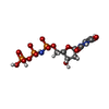  Mass: 467.157 Da / Num. of mol.: 1 / Source method: obtained synthetically / Formula: C9H16N3O13P3 Mass: 467.157 Da / Num. of mol.: 1 / Source method: obtained synthetically / Formula: C9H16N3O13P3 |
| #4: Chemical | ChemComp-TRS /   Mass: 122.143 Da / Num. of mol.: 1 / Source method: obtained synthetically / Formula: C4H12NO3 / Comment: pH buffer*YM Mass: 122.143 Da / Num. of mol.: 1 / Source method: obtained synthetically / Formula: C4H12NO3 / Comment: pH buffer*YM |
| #5: Water | ChemComp-HOH /  Mass: 18.015 Da / Num. of mol.: 132 / Source method: isolated from a natural source / Formula: H2O Mass: 18.015 Da / Num. of mol.: 132 / Source method: isolated from a natural source / Formula: H2O |
-Experimental details
-Experiment
| Experiment | Method: X-RAY DIFFRACTION / Number of used crystals: 1 |
|---|
- Sample preparation
| Crystal | Density Matthews: 1.98 Å3/Da / Density % sol: 38.02 % |
|---|---|
| Crystal grow | Temperature: 298 K / Method: vapor diffusion, hanging drop / pH: 8.5 Details: 1.5M ammonium sulfate, 0.1M Tris/HCl, 12% glycerol, pH 8.5, VAPOR DIFFUSION, HANGING DROP, temperature 298K |
-Data collection
| Diffraction | Mean temperature: 100 K |
|---|---|
| Diffraction source | Source: ROTATING ANODE / Type: RIGAKU / Wavelength: 1.5418 Å |
| Detector | Type: RIGAKU RAXIS II / Detector: IMAGE PLATE |
| Radiation | Monochromator: GRAPHITE / Protocol: SINGLE WAVELENGTH / Monochromatic (M) / Laue (L): M / Scattering type: x-ray |
| Radiation wavelength | Wavelength: 1.5418 Å / Relative weight: 1 |
| Reflection | Resolution: 1.49→20 Å / Num. all: 21589 / Num. obs: 21589 / % possible obs: 94.1 % / Observed criterion σ(I): -3 / Redundancy: 3.95 % / Biso Wilson estimate: 19.3 Å2 / Rsym value: 4.1 / Net I/σ(I): 24.57 |
| Reflection shell | Resolution: 1.49→1.58 Å / Redundancy: 2.3 % / Mean I/σ(I) obs: 3.3 / Num. unique all: 2595 / Rsym value: 34.8 / % possible all: 70.5 |
- Processing
| Software |
| ||||||||||||||||||||||||||||||||||||||||||||||||||||||||||||||||||||||||||||||||||||||||||
|---|---|---|---|---|---|---|---|---|---|---|---|---|---|---|---|---|---|---|---|---|---|---|---|---|---|---|---|---|---|---|---|---|---|---|---|---|---|---|---|---|---|---|---|---|---|---|---|---|---|---|---|---|---|---|---|---|---|---|---|---|---|---|---|---|---|---|---|---|---|---|---|---|---|---|---|---|---|---|---|---|---|---|---|---|---|---|---|---|---|---|---|
| Refinement | Method to determine structure: MOLECULAR REPLACEMENT Starting model: pdb entry 1MQ7 Resolution: 1.49→19.44 Å / Cor.coef. Fo:Fc: 0.972 / Cor.coef. Fo:Fc free: 0.96 / SU B: 2.184 / SU ML: 0.045 / TLS residual ADP flag: LIKELY RESIDUAL / Isotropic thermal model: TLS / Cross valid method: THROUGHOUT / ESU R: 0.063 / ESU R Free: 0.065 / Stereochemistry target values: MAXIMUM LIKELIHOOD / Details: HYDROGENS HAVE BEEN ADDED IN THE RIDING POSITIONS
| ||||||||||||||||||||||||||||||||||||||||||||||||||||||||||||||||||||||||||||||||||||||||||
| Solvent computation | Ion probe radii: 0.8 Å / Shrinkage radii: 0.8 Å / VDW probe radii: 1.2 Å / Solvent model: BABINET MODEL WITH MASK | ||||||||||||||||||||||||||||||||||||||||||||||||||||||||||||||||||||||||||||||||||||||||||
| Displacement parameters | Biso mean: 18.838 Å2
| ||||||||||||||||||||||||||||||||||||||||||||||||||||||||||||||||||||||||||||||||||||||||||
| Refine analyze | Luzzati coordinate error obs: 0.141 Å | ||||||||||||||||||||||||||||||||||||||||||||||||||||||||||||||||||||||||||||||||||||||||||
| Refinement step | Cycle: LAST / Resolution: 1.49→19.44 Å
| ||||||||||||||||||||||||||||||||||||||||||||||||||||||||||||||||||||||||||||||||||||||||||
| Refine LS restraints |
| ||||||||||||||||||||||||||||||||||||||||||||||||||||||||||||||||||||||||||||||||||||||||||
| LS refinement shell | Resolution: 1.49→1.529 Å / Total num. of bins used: 20
| ||||||||||||||||||||||||||||||||||||||||||||||||||||||||||||||||||||||||||||||||||||||||||
| Refinement TLS params. | Method: refined / Origin x: 20.85 Å / Origin y: 17.7538 Å / Origin z: 39.5449 Å
|
