 Movie
Movie Controller

[English] 日本語
 Yorodumi
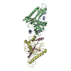
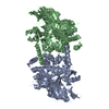
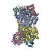
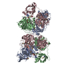
- PDB-1ibw: STRUCTURE OF THE D53,54N MUTANT OF HISTIDINE DECARBOXYLASE BOUND ... -
+ Open data

- Basic information
| Entry | Database: PDB / ID: 1ibw | ||||||||||||
|---|---|---|---|---|---|---|---|---|---|---|---|---|---|
| Title | STRUCTURE OF THE D53,54N MUTANT OF HISTIDINE DECARBOXYLASE BOUND WITH HISTIDINE METHYL ESTER AT 25 C | ||||||||||||
 Components Components |
| ||||||||||||
 Keywords Keywords | LYASE / SUBSTRATE-INDUCED ACTIVATION / ACTIVE FORM / SITE-DIRECTED MUTANT / PYRUVOYL / CARBOXY-LYASE | ||||||||||||
| Function / homology |  Function and homology information Function and homology informationhistidine decarboxylase / histidine decarboxylase activity / L-histidine metabolic process Similarity search - Function | ||||||||||||
| Biological species |  Lactobacillus sp. 30A (bacteria) Lactobacillus sp. 30A (bacteria) | ||||||||||||
| Method |  X-RAY DIFFRACTION / MOLECULAR REPLACEMENT / Resolution: 3.2 Å X-RAY DIFFRACTION / MOLECULAR REPLACEMENT / Resolution: 3.2 Å | ||||||||||||
 Authors Authors | Worley, S. / Schelp, E. / Monzingo, A.F. / Ernst, S. / Robertus, J.D. | ||||||||||||
 Citation Citation | Journal: Proteins / Year: 2002 Title: Structure and cooperativity of a T-state mutant of histidine decarboxylase from Lactobacillus 30a. Authors: Worley, S. / Schelp, E. / Monzingo, A.F. / Ernst, S. / Robertus, J.D. | ||||||||||||
| History |
|
- Structure visualization
| Structure viewer | Molecule: MolmilJmol/JSmol |
|---|
- Downloads & links
-Download
| PDBx/mmCIF format | 1ibw.cif.gz | 187.4 KB | Display | PDBx/mmCIF format |
|---|---|---|---|---|
| PDB format | pdb1ibw.ent.gz | 150.9 KB | Display | PDB format |
| PDBx/mmJSON format | 1ibw.json.gz | Tree view | PDBx/mmJSON format | |
| Others |  Other downloads Other downloads |
-Validation report
| Summary document | 1ibw_validation.pdf.gz | 484.9 KB | Display | wwPDB validaton report |
|---|---|---|---|---|
| Full document | 1ibw_full_validation.pdf.gz | 508.2 KB | Display | |
| Data in XML | 1ibw_validation.xml.gz | 36.3 KB | Display | |
| Data in CIF | 1ibw_validation.cif.gz | 48 KB | Display | |
| Arichive directory | https://data.pdbj.org/pub/pdb/validation_reports/ib/1ibwftp://data.pdbj.org/pub/pdb/validation_reports/ib/1ibw | HTTPS FTP |
-Related structure data
| Related structure data | 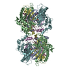 1ibtC 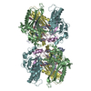 1ibuC 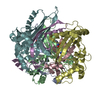 1ibvC 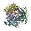 1pyaS S: Starting model for refinement C: citing same article ( |
|---|---|
| Similar structure data |


-Links
 PDBj
PDBj- Assembly
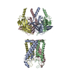
| Deposited unit | 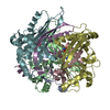
| ||||||||
|---|---|---|---|---|---|---|---|---|---|
| 1 | 
| ||||||||
| Unit cell |
| ||||||||
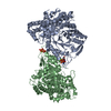
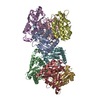
| Details | The biological assembly is a hexamer generated by applying a crystallographic two-fold to the trimer in the asymmetric unit: -x, y, -z + 1/2 |
-Components
| #1: Protein | Mass: 8848.862 Da / Num. of mol.: 3 / Fragment: BETA CHAIN (RESIDUES 1-81) / Mutation: D53N, D54N Source method: isolated from a genetically manipulated source Source: (gene. exp.) Lactobacillus sp. 30A (bacteria) / Strain: 30A / Gene: HDCA / Production host: #2: Protein | Mass: 25285.375 Da / Num. of mol.: 3 / Fragment: ALPHA CHAIN (RESIDUES 82-310) Source method: isolated from a genetically manipulated source Source: (gene. exp.) Lactobacillus sp. 30A (bacteria) / Strain: 30A / Gene: hdcA / Production host: #3: Chemical | 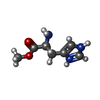  Type: L-peptide linking / Mass: 170.189 Da / Num. of mol.: 3 / Source method: obtained synthetically / Formula: C7H12N3O2 / Comment: inhibitor*YM Type: L-peptide linking / Mass: 170.189 Da / Num. of mol.: 3 / Source method: obtained synthetically / Formula: C7H12N3O2 / Comment: inhibitor*YM |
|---|
-Experimental details
-Experiment
| Experiment | Method: X-RAY DIFFRACTION / Number of used crystals: 1 |
|---|
- Sample preparation
| Crystal | Density Matthews: 2.87 Å3/Da / Density % sol: 57.14 % | ||||||||||||||||||||||||||||||
|---|---|---|---|---|---|---|---|---|---|---|---|---|---|---|---|---|---|---|---|---|---|---|---|---|---|---|---|---|---|---|---|
| Crystal grow | Temperature: 298 K / Method: vapor diffusion, hanging drop / pH: 4.6 Details: PEG 400, PEG 4000, sodium acetate, pH 4.6, VAPOR DIFFUSION, HANGING DROP, temperature 298K | ||||||||||||||||||||||||||||||
| Crystal grow | *PLUS | ||||||||||||||||||||||||||||||
| Components of the solutions | *PLUS
|
-Data collection
| Diffraction | Mean temperature: 298 K |
|---|---|
| Diffraction source | Source: ROTATING ANODE / Type: RIGAKU RU200 / Wavelength: 1.5418 |
| Detector | Type: RIGAKU RAXIS IV / Detector: IMAGE PLATE / Date: Jun 21, 2000 |
| Radiation | Monochromator: DOUBLE FOCUSSING MIRRORS (NI & PT) + NI FILTER Protocol: SINGLE WAVELENGTH / Monochromatic (M) / Laue (L): M / Scattering type: x-ray |
| Radiation wavelength | Wavelength: 1.5418 Å / Relative weight: 1 |
| Reflection | Resolution: 3.2→20 Å / Num. all: 15945 / Num. obs: 15945 / % possible obs: 80.7 % / Observed criterion σ(I): 0 / Redundancy: 1.8 % / Biso Wilson estimate: 66.8 Å2 / Rmerge(I) obs: 0.103 / Net I/σ(I): 10.7 |
| Reflection shell | Resolution: 3.2→3.31 Å / Redundancy: 1.8 % / Rmerge(I) obs: 0.454 / % possible all: 84.8 |
| Reflection | *PLUS Highest resolution: 3.2 Å / Lowest resolution: 20 Å / Redundancy: 1.8 % |
| Reflection shell | *PLUS Mean I/σ(I) obs: 2.1 |
- Processing
| Software |
| ||||||||||||||||||||
|---|---|---|---|---|---|---|---|---|---|---|---|---|---|---|---|---|---|---|---|---|---|
| Refinement | Method to determine structure: MOLECULAR REPLACEMENT Starting model: PDB ENTRY 1PYA Resolution: 3.2→20 Å / Cross valid method: THROUGHOUT / σ(F): 2 / Stereochemistry target values: ENGH & HUBER
| ||||||||||||||||||||
| Refinement step | Cycle: LAST / Resolution: 3.2→20 Å
| ||||||||||||||||||||
| Refine LS restraints |
| ||||||||||||||||||||
| Refinement | *PLUS σ(F): 2 | ||||||||||||||||||||
| Solvent computation | *PLUS | ||||||||||||||||||||
| Displacement parameters | *PLUS |
