 Movie
Movie Controller


+ Open data

- Basic information
| Entry | Database: PDB / ID: 1ibh | ||||||
|---|---|---|---|---|---|---|---|
| Title | X-RAY 3D STRUCTURE OF P.LEIOGNATHI CU,ZN SOD MUTANT M41I | ||||||
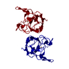
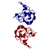
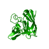
 Components Components | CU,ZN SUPEROXIDE DISMUTASE | ||||||
 Keywords Keywords | OXIDOREDUCTASE / prokaryotic superoxide dismutase / subunit interaction | ||||||
| Function / homology |  Function and homology information Function and homology informationsuperoxide dismutase / superoxide dismutase activity / periplasmic space / copper ion binding Similarity search - Function | ||||||
| Biological species |  Photobacterium leiognathi (bacteria) Photobacterium leiognathi (bacteria) | ||||||
| Method |  X-RAY DIFFRACTION / SYNCHROTRON / FOURIER SYNTHESIS / Resolution: 2 Å X-RAY DIFFRACTION / SYNCHROTRON / FOURIER SYNTHESIS / Resolution: 2 Å | ||||||
 Authors Authors | Stroppolo, M.E. / Pesce, A. / D'Orazio, M. / O'Neill, P. / Bordo, D. / Rosano, C. / Milani, M. / Battistoni, A. / Bolognesi, M. / Desideri, A. | ||||||
 Citation Citation | Journal: J.Mol.Biol. / Year: 2001 Title: Single mutations at the subunit interface modulate copper reactivity in Photobacterium leiognathi Cu,Zn superoxide dismutase. Authors: Stroppolo, M.E. / Pesce, A. / D'Orazio, M. / O'Neill, P. / Bordo, D. / Rosano, C. / Milani, M. / Battistoni, A. / Bolognesi, M. / Desideri, A. | ||||||
| History |
|
- Structure visualization
| Structure viewer | Molecule: MolmilJmol/JSmol |
|---|
- Downloads & links
-Download
| PDBx/mmCIF format | 1ibh.cif.gz | 42.4 KB | Display | PDBx/mmCIF format |
|---|---|---|---|---|
| PDB format | pdb1ibh.ent.gz | 29.3 KB | Display | PDB format |
| PDBx/mmJSON format | 1ibh.json.gz | Tree view | PDBx/mmJSON format | |
| Others |  Other downloads Other downloads |
-Validation report
| Arichive directory | https://data.pdbj.org/pub/pdb/validation_reports/ib/1ibhftp://data.pdbj.org/pub/pdb/validation_reports/ib/1ibh | HTTPS FTP |
|---|
-Related structure data


-Links
 PDBj
PDBj
- Assembly
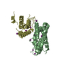
| Deposited unit | 
| ||||||||
|---|---|---|---|---|---|---|---|---|---|
| 1 | 
| ||||||||
| Unit cell |
|
-Components
| #1: Protein | Mass: 15795.815 Da / Num. of mol.: 1 / Mutation: M41I Source method: isolated from a genetically manipulated source Source: (gene. exp.) Photobacterium leiognathi (bacteria) / Production host: |
|---|---|
| #2: Chemical | ChemComp-ZN /   Mass: 65.409 Da / Num. of mol.: 1 / Source method: obtained synthetically / Formula: Zn Mass: 65.409 Da / Num. of mol.: 1 / Source method: obtained synthetically / Formula: Zn |
| #3: Chemical | ChemComp-CU /   Mass: 63.546 Da / Num. of mol.: 1 / Source method: obtained synthetically / Formula: Cu Mass: 63.546 Da / Num. of mol.: 1 / Source method: obtained synthetically / Formula: Cu |
| #4: Water | ChemComp-HOH /  Mass: 18.015 Da / Num. of mol.: 73 / Source method: isolated from a natural source / Formula: H2O Mass: 18.015 Da / Num. of mol.: 73 / Source method: isolated from a natural source / Formula: H2O |
| Has protein modification | Y |
-Experimental details
-Experiment
| Experiment | Method: X-RAY DIFFRACTION / Number of used crystals: 1 |
|---|
- Sample preparation
| Crystal | Density Matthews: 2.21 Å3/Da / Density % sol: 44.35 % | ||||||||||||||||||||||||||||||
|---|---|---|---|---|---|---|---|---|---|---|---|---|---|---|---|---|---|---|---|---|---|---|---|---|---|---|---|---|---|---|---|
| Crystal grow | Temperature: 301 K / Method: vapor diffusion, hanging drop / pH: 4.5 Details: PEG 8k, NaCl, pH 4.5, VAPOR DIFFUSION, HANGING DROP, temperature 301K | ||||||||||||||||||||||||||||||
| Crystal grow | *PLUS Temperature: 100 K / Method: unknown | ||||||||||||||||||||||||||||||
| Components of the solutions | *PLUS
|
-Data collection
| Diffraction | Mean temperature: 100 K |
|---|---|
| Diffraction source | Source: SYNCHROTRON / Site: EMBL/DESY, HAMBURG  / Beamline: X11 / Wavelength: 1 Å / Beamline: X11 / Wavelength: 1 Å |
| Detector | Type: MARRESEARCH / Detector: IMAGE PLATE / Date: Jul 2, 1999 / Details: mirror |
| Radiation | Protocol: SINGLE WAVELENGTH / Monochromatic (M) / Laue (L): M / Scattering type: x-ray |
| Radiation wavelength | Wavelength: 1 Å / Relative weight: 1 |
| Reflection | Resolution: 2→18 Å / Num. all: 9449 / Num. obs: 9449 / % possible obs: 99.6 % / Observed criterion σ(F): 0 / Observed criterion σ(I): 0 / Redundancy: 11.3 % / Biso Wilson estimate: 30 Å2 / Rmerge(I) obs: 0.063 / Rsym value: 0.063 / Net I/σ(I): 26 |
| Reflection | *PLUS Lowest resolution: 18 Å / Num. measured all: 106644 |
- Processing
| Software |
| |||||||||||||||||||||||||
|---|---|---|---|---|---|---|---|---|---|---|---|---|---|---|---|---|---|---|---|---|---|---|---|---|---|---|
| Refinement | Method to determine structure: FOURIER SYNTHESIS Starting model: P.leiognathi Cu,Zn SOD wild type Resolution: 2→18 Å / σ(F): 0 / σ(I): 0 / Stereochemistry target values: Engh & Huber
| |||||||||||||||||||||||||
| Refinement step | Cycle: LAST / Resolution: 2→18 Å
| |||||||||||||||||||||||||
| Refine LS restraints |
| |||||||||||||||||||||||||
| Software | *PLUS Name: CNS / Version: 0.9 / Classification: refinement | |||||||||||||||||||||||||
| Refinement | *PLUS Highest resolution: 2 Å / Lowest resolution: 18 Å / σ(F): 0 / % reflection Rfree: 10 % / Rfactor obs: 0.229 | |||||||||||||||||||||||||
| Solvent computation | *PLUS | |||||||||||||||||||||||||
| Displacement parameters | *PLUS Biso mean: 27 Å2 |
