 Movie
Movie Controller


+ Open data

- Basic information
| Entry | Database: PDB / ID: 1yai | ||||||
|---|---|---|---|---|---|---|---|
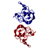
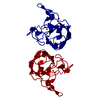
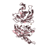
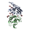
| Title | X-RAY STRUCTURE OF A BACTERIAL COPPER,ZINC SUPEROXIDE DISMUTASE | ||||||
 Components Components | COPPER, ZINC SUPEROXIDE DISMUTASE | ||||||
 Keywords Keywords | OXIDOREDUCTASE / BETA-BARREL / METALLOENZYME / MACROMOLECULAR ASSEMBLY | ||||||
| Function / homology |  Function and homology information Function and homology informationsuperoxide dismutase / superoxide dismutase activity / periplasmic space / copper ion binding Similarity search - Function | ||||||
| Biological species |  Photobacterium leiognathi (bacteria) Photobacterium leiognathi (bacteria) | ||||||
| Method |  X-RAY DIFFRACTION / MOLECULAR REPLACEMENT / Resolution: 1.9 Å X-RAY DIFFRACTION / MOLECULAR REPLACEMENT / Resolution: 1.9 Å | ||||||
 Authors Authors | Bourne, Y. / Redford, S.M. / Lo, T.P. / Tainer, J.A. / Getzoff, E.D. | ||||||
 Citation Citation | Journal: Proc.Natl.Acad.Sci.USA / Year: 1996 Title: Novel dimeric interface and electrostatic recognition in bacterial Cu,Zn superoxide dismutase. Authors: Bourne, Y. / Redford, S.M. / Steinman, H.M. / Lepock, J.R. / Tainer, J.A. / Getzoff, E.D. #1: Journal: J.Mol.Biol. / Year: 1990Title: Crystallographic Characterization of a Cu,Zn Superoxide Dismutase from Photobacterium Leiognathi Authors: Redford, S.M. / Mcree, D.E. / Getzoff, E.D. / Steinman, H.M. / Tainer, J.A. | ||||||
| History |
|
- Structure visualization
| Structure viewer | Molecule: MolmilJmol/JSmol |
|---|
- Downloads & links
-Download
| PDBx/mmCIF format | 1yai.cif.gz | 102.7 KB | Display | PDBx/mmCIF format |
|---|---|---|---|---|
| PDB format | pdb1yai.ent.gz | 78.5 KB | Display | PDB format |
| PDBx/mmJSON format | 1yai.json.gz | Tree view | PDBx/mmJSON format | |
| Others |  Other downloads Other downloads |
-Validation report
| Arichive directory | https://data.pdbj.org/pub/pdb/validation_reports/ya/1yaiftp://data.pdbj.org/pub/pdb/validation_reports/ya/1yai | HTTPS FTP |
|---|
-Related structure data
| Related structure data |  3sodS S: Starting model for refinement |
|---|---|
| Similar structure data |


-Links
 PDBj
PDBj
- Assembly
| Deposited unit | 
| ||||||||
|---|---|---|---|---|---|---|---|---|---|
| 1 | 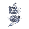
| ||||||||
| 2 | 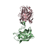
| ||||||||
| Unit cell |
| ||||||||
| Noncrystallographic symmetry (NCS) | NCS oper: (Code: given Matrix: (-0.20969, -0.65398, -0.72687), Vector: |
-Components
| #1: Protein | Mass: 15801.800 Da / Num. of mol.: 3 Source method: isolated from a genetically manipulated source Source: (gene. exp.) Photobacterium leiognathi (bacteria) / Strain: MC1061 / Gene: PHSOD / Plasmid: PPHSOD1LACI / Gene (production host): PHSOD / Production host: #2: Chemical |   Mass: 63.546 Da / Num. of mol.: 3 / Source method: obtained synthetically / Formula: Cu Mass: 63.546 Da / Num. of mol.: 3 / Source method: obtained synthetically / Formula: Cu#3: Chemical |   Mass: 65.409 Da / Num. of mol.: 3 / Source method: obtained synthetically / Formula: Zn Mass: 65.409 Da / Num. of mol.: 3 / Source method: obtained synthetically / Formula: Zn#4: Water | ChemComp-HOH / |  Mass: 18.015 Da / Num. of mol.: 358 / Source method: isolated from a natural source / Formula: H2O Mass: 18.015 Da / Num. of mol.: 358 / Source method: isolated from a natural source / Formula: H2OCompound details | IN ALL THREE CHAINS, ARG 144 IS THE CATALYTICALLY IMPORTANT RESIDUE FOR SUPEROXIDE ANION ...IN ALL THREE CHAINS, ARG 144 IS THE CATALYTICA | Has protein modification | Y | |
|---|
-Experimental details
-Experiment
| Experiment | Method: X-RAY DIFFRACTION / Number of used crystals: 1 |
|---|
- Sample preparation
| Crystal | Density Matthews: 2.41 Å3/Da / Density % sol: 48 % | |||||||||||||||||||||||||
|---|---|---|---|---|---|---|---|---|---|---|---|---|---|---|---|---|---|---|---|---|---|---|---|---|---|---|
| Crystal grow | pH: 6.5 / Details: 42% MPD, 60 MM POTASSIUM PHOSPHATE, PH 6.5 | |||||||||||||||||||||||||
| Crystal grow | *PLUS Temperature: 20 ℃ / Method: vapor diffusion / Details: macroseeding | |||||||||||||||||||||||||
| Components of the solutions | *PLUS
|
-Data collection
| Diffraction | Mean temperature: 293 K |
|---|---|
| Diffraction source | Source: ROTATING ANODE / Type: RIGAKU / Wavelength: 1.5418 |
| Detector | Type: SIEMENS / Detector: AREA DETECTOR / Date: Jul 20, 1991 |
| Radiation | Monochromator: NI FILTER / Monochromatic (M) / Laue (L): M / Scattering type: x-ray |
| Radiation wavelength | Wavelength: 1.5418 Å / Relative weight: 1 |
| Reflection | Highest resolution: 1.9 Å / Num. obs: 116490 / % possible obs: 83 % / Observed criterion σ(I): 0 / Redundancy: 4 % / Rsym value: 0.049 |
| Reflection | *PLUS Num. obs: 30277 / Num. measured all: 116490 / Rmerge(I) obs: 0.049 |
- Processing
| Software |
| ||||||||||||||||||||||||||||||||||||||||||||||||||
|---|---|---|---|---|---|---|---|---|---|---|---|---|---|---|---|---|---|---|---|---|---|---|---|---|---|---|---|---|---|---|---|---|---|---|---|---|---|---|---|---|---|---|---|---|---|---|---|---|---|---|---|
| Refinement | Method to determine structure: MOLECULAR REPLACEMENT Starting model: PDB ENTRY 3SOD Resolution: 1.9→20 Å / Isotropic thermal model: TNT BCORREL / σ(F): 0 / Stereochemistry target values: TNT PROTGEO / Details: X-PLOR ALSO USED
| ||||||||||||||||||||||||||||||||||||||||||||||||||
| Solvent computation | Solvent model: BABINET SCALING / Bsol: 204 Å2 / ksol: 0.65 e/Å3 | ||||||||||||||||||||||||||||||||||||||||||||||||||
| Refinement step | Cycle: LAST / Resolution: 1.9→20 Å
| ||||||||||||||||||||||||||||||||||||||||||||||||||
| Refine LS restraints |
| ||||||||||||||||||||||||||||||||||||||||||||||||||
| Software | *PLUS Name: TNT / Version: 5EB / Classification: refinement | ||||||||||||||||||||||||||||||||||||||||||||||||||
| Refinement | *PLUS Rfactor obs: 0.149 | ||||||||||||||||||||||||||||||||||||||||||||||||||
| Solvent computation | *PLUS | ||||||||||||||||||||||||||||||||||||||||||||||||||
| Displacement parameters | *PLUS | ||||||||||||||||||||||||||||||||||||||||||||||||||
| Refine LS restraints | *PLUS
|
