 Movie
Movie Controller

[English] 日本語
 Yorodumi
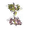
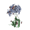
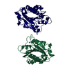
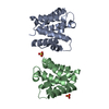
- PDB-1bzo: THREE-DIMENSIONAL STRUCTURE OF PROKARYOTIC CU,ZN SUPEROXIDE DISMU... -
+ Open data

- Basic information
| Entry | Database: PDB / ID: 1bzo | ||||||
|---|---|---|---|---|---|---|---|
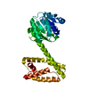
| Title | THREE-DIMENSIONAL STRUCTURE OF PROKARYOTIC CU,ZN SUPEROXIDE DISMUTASE FROM P.LEIOGNATHI, SOLVED BY X-RAY CRYSTALLOGRAPHY. | ||||||
 Components Components | PROTEIN (SUPEROXIDE DISMUTASE) | ||||||
 Keywords Keywords | OXIDOREDUCTASE / MONOMERIC CU / ZN SUPEROXIDE DISMUTASE / PROTEIN-SUBUNIT RECOGNITION / PROTEIN ELECTROSTATIC | ||||||
| Function / homology |  Function and homology information Function and homology informationsuperoxide dismutase / superoxide dismutase activity / periplasmic space / copper ion binding Similarity search - Function | ||||||
| Biological species |  Photobacterium leiognathi (bacteria) Photobacterium leiognathi (bacteria) | ||||||
| Method |  X-RAY DIFFRACTION / SYNCHROTRON / MOLECULAR REPLACEMENT / Resolution: 2.1 Å X-RAY DIFFRACTION / SYNCHROTRON / MOLECULAR REPLACEMENT / Resolution: 2.1 Å | ||||||
 Authors Authors | Bordo, D. / Matak, D. / Djinovic-Carugo, K. / Rosano, C. / Pesce, A. / Bolognesi, M. / Stroppolo, M.E. / Falconi, M. / Battistoni, A. / Desideri, A. | ||||||
 Citation Citation | Journal: J.Mol.Biol. / Year: 1999 Title: Evolutionary constraints for dimer formation in prokaryotic Cu,Zn superoxide dismutase. Authors: Bordo, D. / Matak, D. / Djinovic-Carugo, K. / Rosano, C. / Pesce, A. / Bolognesi, M. / Stroppolo, M.E. / Falconi, M. / Battistoni, A. / Desideri, A. #1: Journal: Proc.Natl.Acad.Sci.USA / Year: 1996Title: Novel Dimeric Interface and Electrostatic Recognition in Bacterial Cu,Zn Superoxide Dismutase Authors: Bourne, Y. / Redford, S.M. / Steinman, H.M. / Lepock, J.R. / Tainer, J.A. / Getzoff, E.D. | ||||||
| History |
|
- Structure visualization
| Structure viewer | Molecule: MolmilJmol/JSmol |
|---|
- Downloads & links
-Download
| PDBx/mmCIF format | 1bzo.cif.gz | 44.9 KB | Display | PDBx/mmCIF format |
|---|---|---|---|---|
| PDB format | pdb1bzo.ent.gz | 31.1 KB | Display | PDB format |
| PDBx/mmJSON format | 1bzo.json.gz | Tree view | PDBx/mmJSON format | |
| Others |  Other downloads Other downloads |
-Validation report
| Arichive directory | https://data.pdbj.org/pub/pdb/validation_reports/bz/1bzoftp://data.pdbj.org/pub/pdb/validation_reports/bz/1bzo | HTTPS FTP |
|---|
-Related structure data
| Similar structure data |
|---|


-Links
 PDBj
PDBj

- Assembly
| Deposited unit | 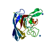
| ||||||||
|---|---|---|---|---|---|---|---|---|---|
| 1 | 
| ||||||||
| Unit cell |
|
-Components
| #1: Protein | Mass: 15813.854 Da / Num. of mol.: 1 Source method: isolated from a genetically manipulated source Source: (gene. exp.) Photobacterium leiognathi (bacteria) / Cellular location: PERIPLASMIC SPACE / Production host: | ||||||||
|---|---|---|---|---|---|---|---|---|---|
| #2: Chemical | ChemComp-ZN /   Mass: 65.409 Da / Num. of mol.: 1 / Source method: obtained synthetically / Formula: Zn Mass: 65.409 Da / Num. of mol.: 1 / Source method: obtained synthetically / Formula: Zn | ||||||||
| #3: Chemical | ChemComp-CU /   Mass: 63.546 Da / Num. of mol.: 1 / Source method: obtained synthetically / Formula: Cu Mass: 63.546 Da / Num. of mol.: 1 / Source method: obtained synthetically / Formula: Cu | ||||||||
| #4: Chemical | 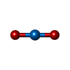  Mass: 270.028 Da / Num. of mol.: 2 / Source method: obtained synthetically / Formula: O2U Mass: 270.028 Da / Num. of mol.: 2 / Source method: obtained synthetically / Formula: O2U#5: Water | ChemComp-HOH / |  Mass: 18.015 Da / Num. of mol.: 102 / Source method: isolated from a natural source / Formula: H2O Mass: 18.015 Da / Num. of mol.: 102 / Source method: isolated from a natural source / Formula: H2OHas protein modification | Y | Nonpolymer details | ACTIVE SITE CATALYTIC ION COORDINATED TO HIS44, HIS46, HIS61 AND HIS118 ACTIVE SITE METAL ION ...ACTIVE SITE CATALYTIC ION COORDINATE | Sequence details | THE PROTEIN USED IS A TRUNCATED FORM, WITHOUT THE FIRST 22 RESIDUES | |
-Experimental details
-Experiment
| Experiment | Method: X-RAY DIFFRACTION / Number of used crystals: 1 |
|---|
- Sample preparation
| Crystal | Density Matthews: 3.4 Å3/Da / Density % sol: 45.88 % | |||||||||||||||||||||||||
|---|---|---|---|---|---|---|---|---|---|---|---|---|---|---|---|---|---|---|---|---|---|---|---|---|---|---|
| Crystal grow | pH: 4 Details: PEG 8,000 25%, NACL 100 MM, SODIUM ACETATE 50 MM, PH 4, T=28C | |||||||||||||||||||||||||
| Crystal | *PLUS Density % sol: 64 % | |||||||||||||||||||||||||
| Crystal grow | *PLUS Temperature: 28 ℃ / Method: vapor diffusion, sitting drop | |||||||||||||||||||||||||
| Components of the solutions | *PLUS
|
-Data collection
| Diffraction | Mean temperature: 100 K |
|---|---|
| Diffraction source | Source: SYNCHROTRON / Site: EMBL/DESY, HAMBURG  / Beamline: X31 / Beamline: X31 |
| Detector | Type: MARRESEARCH / Detector: IMAGE PLATE / Date: Jun 2, 1997 |
| Radiation | Protocol: SINGLE WAVELENGTH / Monochromatic (M) / Laue (L): M / Scattering type: x-ray |
| Radiation wavelength | Relative weight: 1 |
| Reflection | Resolution: 2.1→24.7 Å / Num. obs: 8469 / % possible obs: 96 % / Redundancy: 3 % / Rmerge(I) obs: 0.06 / Rsym value: 0.09 |
- Processing
| Software |
| ||||||||||||||||||||||||||||||||||||||||||||||||||
|---|---|---|---|---|---|---|---|---|---|---|---|---|---|---|---|---|---|---|---|---|---|---|---|---|---|---|---|---|---|---|---|---|---|---|---|---|---|---|---|---|---|---|---|---|---|---|---|---|---|---|---|
| Refinement | Method to determine structure: MOLECULAR REPLACEMENT Starting model: PREVIOUS MODEL OF THE PROTEIN IN SPACE GROUP C2 NOT DEPOSITED IN PDB Resolution: 2.1→24.7 Å / Isotropic thermal model: TNT BCORREL / σ(F): 0 / Stereochemistry target values: TNT PROTGEO /
| ||||||||||||||||||||||||||||||||||||||||||||||||||
| Solvent computation | Bsol: 206 Å2 / ksol: 0.77 e/Å3 | ||||||||||||||||||||||||||||||||||||||||||||||||||
| Refinement step | Cycle: LAST / Resolution: 2.1→24.7 Å
| ||||||||||||||||||||||||||||||||||||||||||||||||||
| Refine LS restraints |
| ||||||||||||||||||||||||||||||||||||||||||||||||||
| Software | *PLUS Name: TNT / Version: 5E / Classification: refinement | ||||||||||||||||||||||||||||||||||||||||||||||||||
| Refinement | *PLUS Rfactor Rfree: 0.26 / Rfactor Rwork: 0.191 | ||||||||||||||||||||||||||||||||||||||||||||||||||
| Solvent computation | *PLUS | ||||||||||||||||||||||||||||||||||||||||||||||||||
| Displacement parameters | *PLUS | ||||||||||||||||||||||||||||||||||||||||||||||||||
| Refine LS restraints | *PLUS Type: t_planar_d / Dev ideal: 0.01 |
