 Movie
Movie Controller

[English] 日本語
 Yorodumi
- PDB-1i9r: STRUCTURE OF CD40L IN COMPLEX WITH THE FAB FRAGMENT OF HUMANIZED ... -
+ Open data

- Basic information
| Entry | Database: PDB / ID: 1i9r | ||||||
|---|---|---|---|---|---|---|---|
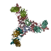
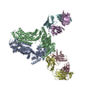
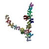
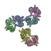
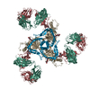
| Title | STRUCTURE OF CD40L IN COMPLEX WITH THE FAB FRAGMENT OF HUMANIZED 5C8 ANTIBODY | ||||||
 Components Components |
| ||||||
 Keywords Keywords | CYTOKINE/IMMUNE SYSTEM / beta-sheet sandwich / immunoglobulin / CYTOKINE-IMMUNE SYSTEM COMPLEX | ||||||
| Function / homology |  Function and homology information Function and homology informationCD40 receptor binding / TNF receptor superfamily (TNFSF) members mediating non-canonical NF-kB pathway / CD40 signaling pathway / regulation of immunoglobulin production / isotype switching / tumor necrosis factor receptor binding / positive regulation of extrinsic apoptotic signaling pathway / leukocyte cell-cell adhesion / positive regulation of interleukin-4 production / B cell proliferation ...CD40 receptor binding / TNF receptor superfamily (TNFSF) members mediating non-canonical NF-kB pathway / CD40 signaling pathway / regulation of immunoglobulin production / isotype switching / tumor necrosis factor receptor binding / positive regulation of extrinsic apoptotic signaling pathway / leukocyte cell-cell adhesion / positive regulation of interleukin-4 production / B cell proliferation / positive regulation of interleukin-10 production / positive regulation of endothelial cell apoptotic process / T cell costimulation / positive regulation of interleukin-12 production / positive regulation of T cell proliferation / B cell differentiation / cytokine activity / : / integrin-mediated signaling pathway / protein serine/threonine kinase activator activity / TNFR2 non-canonical NF-kB pathway / platelet activation / integrin binding / Immunoregulatory interactions between a Lymphoid and a non-Lymphoid cell / positive regulation of canonical NF-kappaB signal transduction / cell surface receptor signaling pathway / inflammatory response / external side of plasma membrane / negative regulation of apoptotic process / cell surface / Golgi apparatus / : / membrane / plasma membrane Similarity search - Function | ||||||
| Biological species |  Homo sapiens (human) Homo sapiens (human) | ||||||
| Method |  X-RAY DIFFRACTION / MOLECULAR REPLACEMENT / Resolution: 3.1 Å X-RAY DIFFRACTION / MOLECULAR REPLACEMENT / Resolution: 3.1 Å | ||||||
 Authors Authors | Karpusas, M. / Lucci, J. / Ferrant, J. / Benjamin, C. / Hsu, Y.-M. | ||||||
 Citation Citation | Journal: Structure / Year: 2001 Title: Structure of CD40 ligand in complex with the Fab fragment of a neutralizing humanized antibody. Authors: Karpusas, M. / Lucci, J. / Ferrant, J. / Benjamin, C. / Taylor, F.R. / Strauch, K. / Garber, E. / Hsu, Y.M. | ||||||
| History |
|
- Structure visualization
| Structure viewer | Molecule: MolmilJmol/JSmol |
|---|
- Downloads & links
-Download
| PDBx/mmCIF format | 1i9r.cif.gz | 329.6 KB | Display | PDBx/mmCIF format |
|---|---|---|---|---|
| PDB format | pdb1i9r.ent.gz | 267 KB | Display | PDB format |
| PDBx/mmJSON format | 1i9r.json.gz | Tree view | PDBx/mmJSON format | |
| Others |  Other downloads Other downloads |
-Validation report
| Arichive directory | https://data.pdbj.org/pub/pdb/validation_reports/i9/1i9rftp://data.pdbj.org/pub/pdb/validation_reports/i9/1i9r | HTTPS FTP |
|---|
-Related structure data
| Related structure data | 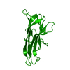 1alyS S: Starting model for refinement |
|---|---|
| Similar structure data |

-Links
 PDBj
PDBj

- Assembly
| Deposited unit | 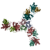
| ||||||||
|---|---|---|---|---|---|---|---|---|---|
| 1 |
| ||||||||
| Unit cell |
|
-Components
| #1: Protein | Mass: 15812.817 Da / Num. of mol.: 3 / Fragment: RESIDUES 116-261 Source method: isolated from a genetically manipulated source Source: (gene. exp.) Homo sapiens (human) / Production host:  Pichia pastoris (fungus) / References: UniProt: P29965 Pichia pastoris (fungus) / References: UniProt: P29965#2: Antibody | Mass: 23304.039 Da / Num. of mol.: 3 / Source method: isolated from a natural source / Source: (natural) Homo sapiens (human) / Cell line: NSO MYELOMA#3: Antibody | Mass: 23881.500 Da / Num. of mol.: 3 / Source method: isolated from a natural source / Source: (natural) Homo sapiens (human) / Cell line: NSO MYELOMA#4: Chemical |   Mass: 65.409 Da / Num. of mol.: 3 / Source method: obtained synthetically / Formula: Zn Mass: 65.409 Da / Num. of mol.: 3 / Source method: obtained synthetically / Formula: ZnHas protein modification | Y | |
|---|
-Experimental details
-Experiment
| Experiment | Method: X-RAY DIFFRACTION / Number of used crystals: 1 |
|---|
- Sample preparation
| Crystal | Density Matthews: 3.1 Å3/Da / Density % sol: 60.7 % | ||||||||||||||||||||||||||||||
|---|---|---|---|---|---|---|---|---|---|---|---|---|---|---|---|---|---|---|---|---|---|---|---|---|---|---|---|---|---|---|---|
| Crystal grow | Temperature: 298 K / Method: vapor diffusion, hanging drop / pH: 6.5 Details: 20% PEG MME 550, 0.1 M MES, 0.01 M zinc sulfate, pH 6.5, VAPOR DIFFUSION, HANGING DROP, temperature 298K | ||||||||||||||||||||||||||||||
| Crystal grow | *PLUS Method: vapor diffusion | ||||||||||||||||||||||||||||||
| Components of the solutions | *PLUS
|
-Data collection
| Diffraction | Mean temperature: 100 K |
|---|---|
| Diffraction source | Source: ROTATING ANODE / Type: RIGAKU RU200 / Wavelength: 1.5418 Å |
| Detector | Type: RIGAKU RAXIS IIC / Detector: IMAGE PLATE / Date: Nov 11, 1999 / Details: mirrors |
| Radiation | Protocol: SINGLE WAVELENGTH / Monochromatic (M) / Laue (L): M / Scattering type: x-ray |
| Radiation wavelength | Wavelength: 1.5418 Å / Relative weight: 1 |
| Reflection | Resolution: 3.1→35 Å / Num. all: 71662 / Num. obs: 46508 / % possible obs: 96.1 % / Observed criterion σ(F): 2 / Observed criterion σ(I): 2 / Rmerge(I) obs: 0.076 / Net I/σ(I): 7.52 |
| Reflection shell | Resolution: 3.1→3.21 Å / Rmerge(I) obs: 0.188 / Mean I/σ(I) obs: 1.97 / % possible all: 87.7 |
| Reflection | *PLUS Lowest resolution: 35 Å |
| Reflection shell | *PLUS % possible obs: 87.7 % |
- Processing
| Software |
| ||||||||||||||||||||
|---|---|---|---|---|---|---|---|---|---|---|---|---|---|---|---|---|---|---|---|---|---|
| Refinement | Method to determine structure: MOLECULAR REPLACEMENT Starting model: pdb entry 1aly Resolution: 3.1→35 Å / Cross valid method: THROUGHOUT / σ(F): 2 / σ(I): 2 / Stereochemistry target values: Engh & Huber Details: For NCS the following 3 chain groups were used: a) A, H, L, b) B,K, X c) C, M, Y. The final rms deviation of main chain atoms between groups is 0.18 Angstrom.
| ||||||||||||||||||||
| Refinement step | Cycle: LAST / Resolution: 3.1→35 Å
| ||||||||||||||||||||
| Refine LS restraints |
| ||||||||||||||||||||
| Refinement | *PLUS Lowest resolution: 35 Å / σ(F): 2 | ||||||||||||||||||||
| Solvent computation | *PLUS | ||||||||||||||||||||
| Displacement parameters | *PLUS |
