 Movie
Movie Controller


+ Open data

- Basic information
| Entry | Database: PDB / ID: 1i5r | ||||||
|---|---|---|---|---|---|---|---|
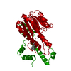
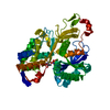
| Title | TYPE 1 17-BETA HYDROXYSTEROID DEHYDROGENASE EM1745 COMPLEX | ||||||
 Components Components | TYPE 1 17 BETA-HYDROXYSTEROID DEHYDROGENASE | ||||||
 Keywords Keywords | OXIDOREDUCTASE / DEHYDROGENASE / 17BETA-HYDROXYSTEROID / HYBRID / INHIBITOR | ||||||
| Function / homology |  Function and homology information Function and homology information17-beta-hydroxysteroid dehydrogenase (NADP+) activity / cellular response to metal ion / estrogen biosynthetic process / 3(or 17)beta-hydroxysteroid dehydrogenase / estradiol binding / Estrogen biosynthesis / testosterone dehydrogenase (NADP+) activity / testosterone biosynthetic process / testosterone dehydrogenase (NAD+) activity / 17beta-estradiol 17-dehydrogenase ...17-beta-hydroxysteroid dehydrogenase (NADP+) activity / cellular response to metal ion / estrogen biosynthetic process / 3(or 17)beta-hydroxysteroid dehydrogenase / estradiol binding / Estrogen biosynthesis / testosterone dehydrogenase (NADP+) activity / testosterone biosynthetic process / testosterone dehydrogenase (NAD+) activity / 17beta-estradiol 17-dehydrogenase / estradiol 17-beta-dehydrogenase [NAD(P)+] activity / NADP+ binding / lysosome organization / The canonical retinoid cycle in rods (twilight vision) / small molecule binding / adipose tissue development / skeletal muscle tissue development / steroid binding / bone development / NADP binding / gene expression / protein homodimerization activity / cytosol Similarity search - Function | ||||||
| Biological species |  Homo sapiens (human) Homo sapiens (human) | ||||||
| Method |  X-RAY DIFFRACTION / SYNCHROTRON / FOURIER SYNTHESIS / Resolution: 1.6 Å X-RAY DIFFRACTION / SYNCHROTRON / FOURIER SYNTHESIS / Resolution: 1.6 Å | ||||||
 Authors Authors | Qiu, W. / Campbell, R.L. / Boivin, P. / Poirier, D. / Lin, S.-X. | ||||||
 Citation Citation | Journal: FASEB J. / Year: 2002 Title: A concerted, rational design of type 1 17beta-hydroxysteroid dehydrogenase inhibitors: estradiol-adenosine hybrids with high affinity Authors: Qiu, W. / Campbell, R.L. / Gangloff, A. / Dupuis, P. / Boivin, R.P. / Tremblay, M.R. / Poirier, D. / Lin, S.X. | ||||||
| History |
|
- Structure visualization
| Structure viewer | Molecule: MolmilJmol/JSmol |
|---|
- Downloads & links
-Download
| PDBx/mmCIF format | 1i5r.cif.gz | 76.4 KB | Display | PDBx/mmCIF format |
|---|---|---|---|---|
| PDB format | pdb1i5r.ent.gz | 54.7 KB | Display | PDB format |
| PDBx/mmJSON format | 1i5r.json.gz | Tree view | PDBx/mmJSON format | |
| Others |  Other downloads Other downloads |
-Validation report
| Arichive directory | https://data.pdbj.org/pub/pdb/validation_reports/i5/1i5rftp://data.pdbj.org/pub/pdb/validation_reports/i5/1i5r | HTTPS FTP |
|---|
-Related structure data
| Related structure data |  1bhsS S: Starting model for refinement |
|---|---|
| Similar structure data |

-Links
 PDBj
PDBj


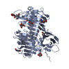
- Assembly
| Deposited unit | 
| ||||||||
|---|---|---|---|---|---|---|---|---|---|
| 1 | 
| ||||||||
| 2 |
| ||||||||
| Unit cell |
|
-Components
| #1: Protein | Mass: 34887.828 Da / Num. of mol.: 1 Source method: isolated from a genetically manipulated source Source: (gene. exp.) Homo sapiens (human) / Production host:   Spodoptera frugiperda (fall armyworm) / Strain (production host): SF9 Spodoptera frugiperda (fall armyworm) / Strain (production host): SF9References: UniProt: P14061, 17beta-estradiol 17-dehydrogenase | ||
|---|---|---|---|
| #2: Chemical | ChemComp-HYC /   Mass: 677.830 Da / Num. of mol.: 1 / Source method: obtained synthetically / Formula: C37H51N5O7 Mass: 677.830 Da / Num. of mol.: 1 / Source method: obtained synthetically / Formula: C37H51N5O7 | ||
| #3: Chemical |   Mass: 92.094 Da / Num. of mol.: 3 / Source method: obtained synthetically / Formula: C3H8O3 Mass: 92.094 Da / Num. of mol.: 3 / Source method: obtained synthetically / Formula: C3H8O3#4: Water | ChemComp-HOH / |  Mass: 18.015 Da / Num. of mol.: 231 / Source method: isolated from a natural source / Formula: H2O Mass: 18.015 Da / Num. of mol.: 231 / Source method: isolated from a natural source / Formula: H2O |
-Experimental details
-Experiment
| Experiment | Method: X-RAY DIFFRACTION / Number of used crystals: 1 |
|---|
- Sample preparation
| Crystal | Density Matthews: 2.12 Å3/Da / Density % sol: 41.85 % | ||||||||||||||||||||||||||||||||||||||||||
|---|---|---|---|---|---|---|---|---|---|---|---|---|---|---|---|---|---|---|---|---|---|---|---|---|---|---|---|---|---|---|---|---|---|---|---|---|---|---|---|---|---|---|---|
| Crystal grow | Temperature: 292 K / Method: vapor diffusion, hanging drop / pH: 7.5 Details: PEG3500, GLYCEROL, SODIUM CHLORIDE, BETA-OCTYL GLUCOSIDE, DITHIOTHREITOL, pH 7.5, VAPOR DIFFUSION, HANGING DROP, temperature 292K | ||||||||||||||||||||||||||||||||||||||||||
| Crystal grow | *PLUS | ||||||||||||||||||||||||||||||||||||||||||
| Components of the solutions | *PLUS
|
-Data collection
| Diffraction | Mean temperature: 100 K |
|---|---|
| Diffraction source | Source: SYNCHROTRON / Site: NSLS  / Beamline: X8C / Wavelength: 1.009 Å / Beamline: X8C / Wavelength: 1.009 Å |
| Detector | Type: ADSC QUANTUM 4 / Detector: CCD / Date: Apr 6, 1999 / Details: parabolic collimating mirror |
| Radiation | Protocol: SINGLE WAVELENGTH / Monochromatic (M) / Laue (L): M / Scattering type: x-ray |
| Radiation wavelength | Wavelength: 1.009 Å / Relative weight: 1 |
| Reflection | Resolution: 1.6→20 Å / Num. all: 155176 / Num. obs: 38831 / % possible obs: 95.2 % / Observed criterion σ(F): 0 / Observed criterion σ(I): 0 / Redundancy: 2.4 % / Biso Wilson estimate: 15.4 Å2 / Rmerge(I) obs: 0.031 / Net I/σ(I): 26.9 |
| Reflection shell | Resolution: 1.6→1.66 Å / Redundancy: 2.2 % / Rmerge(I) obs: 0.109 / Mean I/σ(I) obs: 6.9 / Num. unique all: 3327 / % possible all: 86.2 |
| Reflection | *PLUS Lowest resolution: 20 Å / Num. measured all: 155176 |
| Reflection shell | *PLUS Highest resolution: 1.6 Å / % possible obs: 86.2 % |
- Processing
| Software |
| ||||||||||||||||||||||||||||||||||||||||||
|---|---|---|---|---|---|---|---|---|---|---|---|---|---|---|---|---|---|---|---|---|---|---|---|---|---|---|---|---|---|---|---|---|---|---|---|---|---|---|---|---|---|---|---|
| Refinement | Method to determine structure: FOURIER SYNTHESIS Starting model: PDB ENTRY 1BHS Resolution: 1.6→20 Å / Isotropic thermal model: Isotropic / Cross valid method: THROUGHOUT / σ(F): 0 / σ(I): 0
| ||||||||||||||||||||||||||||||||||||||||||
| Refinement step | Cycle: LAST / Resolution: 1.6→20 Å
| ||||||||||||||||||||||||||||||||||||||||||
| Refine LS restraints |
| ||||||||||||||||||||||||||||||||||||||||||
| LS refinement shell |
| ||||||||||||||||||||||||||||||||||||||||||
| Refinement | *PLUS Lowest resolution: 20 Å / % reflection Rfree: 5 % | ||||||||||||||||||||||||||||||||||||||||||
| Solvent computation | *PLUS | ||||||||||||||||||||||||||||||||||||||||||
| Displacement parameters | *PLUS | ||||||||||||||||||||||||||||||||||||||||||
| Refine LS restraints | *PLUS
|
