 Movie
Movie Controller


+ Open data

- Basic information
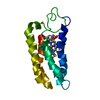
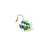
| Entry | Database: PDB / ID: 1i07 | ||||||
|---|---|---|---|---|---|---|---|
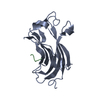
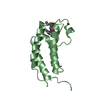
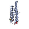
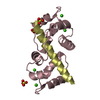
| Title | EPS8 SH3 DOMAIN INTERTWINED DIMER | ||||||
 Components Components | EPIDERMAL GROWTH FACTOR RECEPTOR KINASE SUBSTRATE EPS8 | ||||||
 Keywords Keywords | HORMONE/GROWTH FACTOR / HORMONE-GROWTH FACTOR complex | ||||||
| Function / homology |  Function and homology information Function and homology informationregulation of actin filament length / actin polymerization-dependent cell motility / dendritic cell migration / stereocilium tip / actin crosslink formation / stereocilium bundle / regulation of Rho protein signal transduction / barbed-end actin filament capping / stereocilium / behavioral response to ethanol ...regulation of actin filament length / actin polymerization-dependent cell motility / dendritic cell migration / stereocilium tip / actin crosslink formation / stereocilium bundle / regulation of Rho protein signal transduction / barbed-end actin filament capping / stereocilium / behavioral response to ethanol / exit from mitosis / NMDA selective glutamate receptor complex / positive regulation of ruffle assembly / Rac protein signal transduction / brush border / actin filament bundle assembly / regulation of postsynaptic membrane neurotransmitter receptor levels / Rho protein signal transduction / adult locomotory behavior / cellular response to leukemia inhibitory factor / small GTPase binding / ruffle membrane / regulation of cell shape / growth cone / actin cytoskeleton organization / actin binding / cell cortex / postsynaptic density / synapse / glutamatergic synapse / plasma membrane / cytosol Similarity search - Function | ||||||
| Biological species |  | ||||||
| Method |  X-RAY DIFFRACTION / MOLECULAR REPLACEMENT / Resolution: 1.8 Å X-RAY DIFFRACTION / MOLECULAR REPLACEMENT / Resolution: 1.8 Å | ||||||
 Authors Authors | Kishan, K.V.R. / Newcomer, M.E. | ||||||
 Citation Citation | Journal: Protein Sci. / Year: 2001 Title: Effect of pH and salt bridges on structural assembly: molecular structures of the monomer and intertwined dimer of the Eps8 SH3 domain. Authors: Kishan, K.V. / Newcomer, M.E. / Rhodes, T.H. / Guilliot, S.D. #1: Journal: Nat.Struct.Biol. / Year: 1997Title: The SH3 Domain of Eps8 Exists as a Novel Intertwined Dimer. Authors: Kishan, K.V. / Scita, G. / Wong, W.T. / Di Fiore, P.P. / Newcomer, M.E. | ||||||
| History |
|
- Structure visualization
| Structure viewer | Molecule: MolmilJmol/JSmol |
|---|
- Downloads & links
-Download
| PDBx/mmCIF format | 1i07.cif.gz | 37.8 KB | Display | PDBx/mmCIF format |
|---|---|---|---|---|
| PDB format | pdb1i07.ent.gz | 25.6 KB | Display | PDB format |
| PDBx/mmJSON format | 1i07.json.gz | Tree view | PDBx/mmJSON format | |
| Others |  Other downloads Other downloads |
-Validation report
| Arichive directory | https://data.pdbj.org/pub/pdb/validation_reports/i0/1i07ftp://data.pdbj.org/pub/pdb/validation_reports/i0/1i07 | HTTPS FTP |
|---|
-Related structure data
| Related structure data | 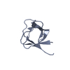 1i0cC 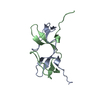 1aojS C: citing same article ( S: Starting model for refinement |
|---|---|
| Similar structure data |


-Links
 PDBj
PDBj
- Assembly
| Deposited unit | 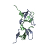
| ||||||||
|---|---|---|---|---|---|---|---|---|---|
| 1 |
| ||||||||
| Unit cell |
|
-Components
| #1: Protein | Mass: 7044.960 Da / Num. of mol.: 2 / Fragment: SH3 DOMAIN Source method: isolated from a genetically manipulated source Source: (gene. exp.)  #2: Water | ChemComp-HOH / |  Mass: 18.015 Da / Num. of mol.: 94 / Source method: isolated from a natural source / Formula: H2O Mass: 18.015 Da / Num. of mol.: 94 / Source method: isolated from a natural source / Formula: H2O |
|---|
-Experimental details
-Experiment
| Experiment | Method: X-RAY DIFFRACTION / Number of used crystals: 1 |
|---|
- Sample preparation
| Crystal | Density Matthews: 1.9 Å3/Da / Density % sol: 35.1 % | ||||||||||||||||||||||||||||||
|---|---|---|---|---|---|---|---|---|---|---|---|---|---|---|---|---|---|---|---|---|---|---|---|---|---|---|---|---|---|---|---|
| Crystal grow | pH: 7 / Details: pH 7.0 | ||||||||||||||||||||||||||||||
| Crystal grow | *PLUS Method: vapor diffusion, hanging drop | ||||||||||||||||||||||||||||||
| Components of the solutions | *PLUS
|
-Data collection
| Diffraction | Mean temperature: 300 K |
|---|---|
| Diffraction source | Source: ROTATING ANODE / Type: RIGAKU RU200 / Wavelength: 1.5418 |
| Detector | Date: Oct 9, 1998 / Details: MSC/YALE MIRRORS |
| Radiation | Protocol: SINGLE WAVELENGTH / Monochromatic (M) / Laue (L): M / Scattering type: x-ray |
| Radiation wavelength | Wavelength: 1.5418 Å / Relative weight: 1 |
| Reflection | Resolution: 1.8→25.93 Å / Num. all: 9088 / Num. obs: 33871 / % possible obs: 94 % / Observed criterion σ(F): 0 / Observed criterion σ(I): 4 / Redundancy: 3.7 % / Biso Wilson estimate: 16.4 Å2 / Limit h max: 15 / Limit h min: -15 / Limit k max: 15 / Limit k min: -15 / Limit l max: 20 / Limit l min: 0 / Observed criterion F max: 370952.57 / Observed criterion F min: 0.3 / Rmerge(I) obs: 0.072 |
| Reflection shell | Resolution: 1.8→1.86 Å / % possible obs: 90.2 % / Rmerge(I) obs: 0.353 / % possible all: 90.2 |
| Reflection | *PLUS Num. obs: 9088 / Num. measured all: 33871 |
- Processing
| Software |
| ||||||||||||||||||||||||||||||||||||||||||||||||||||||||||||||||||||||||||||||||||||||||||
|---|---|---|---|---|---|---|---|---|---|---|---|---|---|---|---|---|---|---|---|---|---|---|---|---|---|---|---|---|---|---|---|---|---|---|---|---|---|---|---|---|---|---|---|---|---|---|---|---|---|---|---|---|---|---|---|---|---|---|---|---|---|---|---|---|---|---|---|---|---|---|---|---|---|---|---|---|---|---|---|---|---|---|---|---|---|---|---|---|---|---|---|
| Refinement | Method to determine structure: MOLECULAR REPLACEMENT Starting model: pdb entry 1AOJ Resolution: 1.8→26 Å / Rfactor Rfree error: 0.01 / Occupancy max: 1 / Occupancy min: 1 / Cross valid method: THROUGHOUT / σ(F): 2 / Stereochemistry target values: Engh & Huber
| ||||||||||||||||||||||||||||||||||||||||||||||||||||||||||||||||||||||||||||||||||||||||||
| Solvent computation | Solvent model: CNS bulk solvent model used / Bsol: 41.5615 Å2 / ksol: 0.307324 e/Å3 | ||||||||||||||||||||||||||||||||||||||||||||||||||||||||||||||||||||||||||||||||||||||||||
| Displacement parameters | Biso max: 51.89 Å2 / Biso mean: 22.36 Å2 / Biso min: 8.75 Å2
| ||||||||||||||||||||||||||||||||||||||||||||||||||||||||||||||||||||||||||||||||||||||||||
| Refine analyze |
| ||||||||||||||||||||||||||||||||||||||||||||||||||||||||||||||||||||||||||||||||||||||||||
| Refinement step | Cycle: LAST / Resolution: 1.8→26 Å
| ||||||||||||||||||||||||||||||||||||||||||||||||||||||||||||||||||||||||||||||||||||||||||
| Refine LS restraints |
| ||||||||||||||||||||||||||||||||||||||||||||||||||||||||||||||||||||||||||||||||||||||||||
| LS refinement shell | Refine-ID: X-RAY DIFFRACTION
| ||||||||||||||||||||||||||||||||||||||||||||||||||||||||||||||||||||||||||||||||||||||||||
| Xplor file |
| ||||||||||||||||||||||||||||||||||||||||||||||||||||||||||||||||||||||||||||||||||||||||||
| Software | *PLUS Name: CNS / Version: 0.5 / Classification: refinement | ||||||||||||||||||||||||||||||||||||||||||||||||||||||||||||||||||||||||||||||||||||||||||
| Refinement | *PLUS Highest resolution: 1.8 Å / Lowest resolution: 26 Å / σ(F): 2 / % reflection Rfree: 6.2 % / Rfactor obs: 0.202 | ||||||||||||||||||||||||||||||||||||||||||||||||||||||||||||||||||||||||||||||||||||||||||
| Solvent computation | *PLUS | ||||||||||||||||||||||||||||||||||||||||||||||||||||||||||||||||||||||||||||||||||||||||||
| Displacement parameters | *PLUS | ||||||||||||||||||||||||||||||||||||||||||||||||||||||||||||||||||||||||||||||||||||||||||
| Refine LS restraints | *PLUS
| ||||||||||||||||||||||||||||||||||||||||||||||||||||||||||||||||||||||||||||||||||||||||||
| LS refinement shell | *PLUS Rfactor Rfree: 0.249 / % reflection Rfree: 4.5 % / Rfactor Rwork: 0.254 |
